

Immature excitatory neurons in the amygdala come of age during puberty
- PMID: 35841648
- PMCID: PMC9289873
- DOI: 10.1016/j.dcn.2022.101133
Immature excitatory neurons in the amygdala come of age during puberty
Abstract
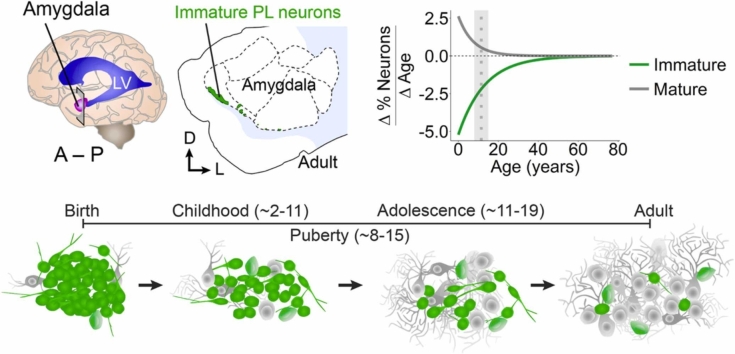
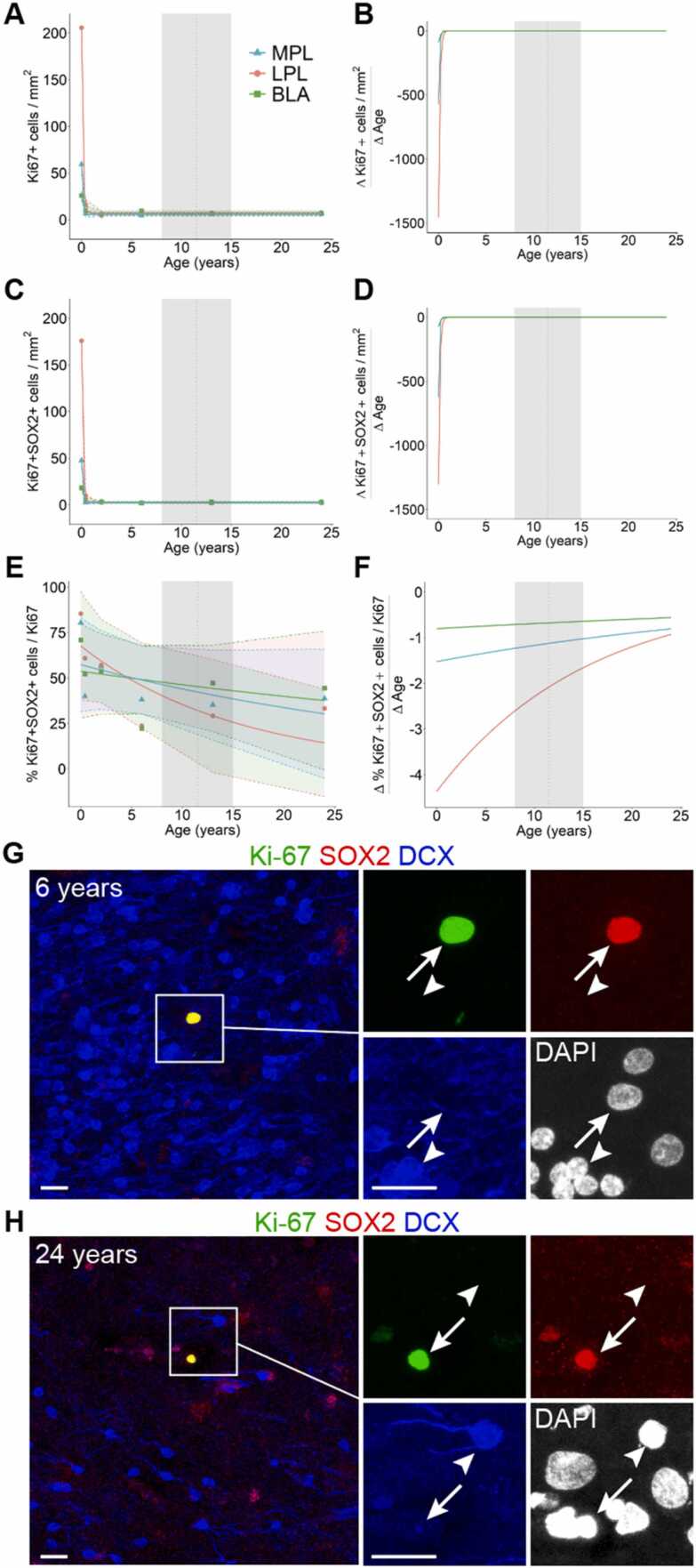
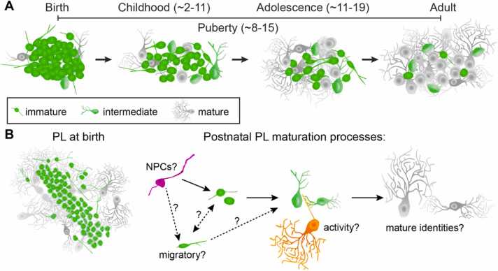
The human amygdala is critical for emotional learning, valence coding, and complex social interactions, all of which mature throughout childhood, puberty, and adolescence. Across these ages, the amygdala paralaminar nucleus (PL) undergoes significant structural changes including increased numbers of mature neurons. The PL contains a large population of immature excitatory neurons at birth, some of which may continue to be born from local progenitors. These progenitors disappear rapidly in infancy, but the immature neurons persist throughout childhood and adolescent ages, indicating that they develop on a protracted timeline. Many of these late-maturing neurons settle locally within the PL, though a small subset appear to migrate into neighboring amygdala subnuclei. Despite its prominent growth during postnatal life and possible contributions to multiple amygdala circuits, the function of the PL remains unknown. PL maturation occurs predominately during late childhood and into puberty when sex hormone levels change. Sex hormones can promote developmental processes such as neuron migration, dendritic outgrowth, and synaptic plasticity, which appear to be ongoing in late-maturing PL neurons. Collectively, we describe how the growth of late-maturing neurons occurs in the right time and place to be relevant for amygdala functions and neuropsychiatric conditions.
Keywords: Amygdala; Development; Migration; Neurogenesis; Paralaminar nucleus; Primates.
Copyright © 2022 The Authors. Published by Elsevier Ltd.. All rights reserved.
Conflict of interest statement
The authors declare that they have no known competing financial interests or personal relationships that could have appeared to influence the work reported in this paper.
Figures
Similar articles
-
Delayed maturation and migration of excitatory neurons in the juvenile mouse paralaminar amygdala.Neuron. 2024 Feb 21;112(4):574-592.e10. doi: 10.1016/j.neuron.2023.11.010. Epub 2023 Dec 11. Neuron. 2024. PMID: 38086370 Free PMC article.
-
Immature excitatory neurons develop during adolescence in the human amygdala.Nat Commun. 2019 Jun 21;10(1):2748. doi: 10.1038/s41467-019-10765-1. Nat Commun. 2019. PMID: 31227709 Free PMC article.
-
Revisiting the hippocampal-amygdala pathway in primates: association with immature-appearing neurons.Neuroscience. 2012 Jun 14;212:104-19. doi: 10.1016/j.neuroscience.2012.03.040. Epub 2012 Apr 19. Neuroscience. 2012. PMID: 22521814 Free PMC article.
-
Where and what is the paralaminar nucleus? A review on a unique and frequently overlooked area of the primate amygdala.Neurosci Biobehav Rev. 2012 Jan;36(1):520-35. doi: 10.1016/j.neubiorev.2011.08.007. Epub 2011 Sep 1. Neurosci Biobehav Rev. 2012. PMID: 21906624 Free PMC article. Review.
-
Postnatal and adult neurogenesis in the development of human disease.Neuroscientist. 2008 Oct;14(5):446-58. doi: 10.1177/1073858408317008. Neuroscientist. 2008. PMID: 18997123 Review.
Cited by
-
Amygdala subregion volumes and apportionment in preadolescents - Associations with age, sex, and body mass index.Dev Cogn Neurosci. 2025 Jun;73:101554. doi: 10.1016/j.dcn.2025.101554. Epub 2025 Mar 20. Dev Cogn Neurosci. 2025. PMID: 40139048 Free PMC article.
-
Microglia Morphology in the Developing Primate Amygdala and Effects of Early Life Stress.eNeuro. 2025 Jan 15;12(1):ENEURO.0466-24.2024. doi: 10.1523/ENEURO.0466-24.2024. Print 2025 Jan. eNeuro. 2025. PMID: 39753372 Free PMC article.
-
Delayed maturation and migration of excitatory neurons in the juvenile mouse paralaminar amygdala.Neuron. 2024 Feb 21;112(4):574-592.e10. doi: 10.1016/j.neuron.2023.11.010. Epub 2023 Dec 11. Neuron. 2024. PMID: 38086370 Free PMC article.
-
Adult sex change leads to extensive forebrain reorganization in clownfish.Biol Sex Differ. 2024 Jul 23;15(1):58. doi: 10.1186/s13293-024-00632-0. Biol Sex Differ. 2024. PMID: 39044232 Free PMC article.
-
Which neurodevelopmental processes continue in humans after birth?Front Neurosci. 2024 Sep 6;18:1434508. doi: 10.3389/fnins.2024.1434508. eCollection 2024. Front Neurosci. 2024. PMID: 39308952 Free PMC article.
References
-
- Amaral D.G., Insausti R. Retrograde transport of D-[3H]-aspartate injected into the monkey amygdaloid complex. Exp. Brain Res. 1992;88:375–388. - PubMed
Publication types
MeSH terms
Grants and funding
LinkOut - more resources
Full Text Sources
