. 2022 Aug 24:786:136800.
doi: 10.1016/j.neulet.2022.136800. Epub 2022 Jul 13.

Changes in somatosensory evoked potentials elicited by lateral cerebellar nucleus deep brain stimulation in the naïve rodent

Affiliations

Changes in somatosensory evoked potentials elicited by lateral cerebellar nucleus deep brain stimulation in the naïve rodent

John K Hermann et al. Neurosci Lett. .

Abstract

Deep brain stimulation (DBS) of the deep cerebellar nuclei has been shown to enhance perilesional cortical excitability and promote motor rehabilitation in preclinical models of cortical ischemia and is currently being evaluated in patients with chronic, post-stroke deficits. Understanding the effects of cerebellar DBS on contralateral sensorimotor cortex may be key to developing approaches to optimize stimulation delivery and treatment outcomes. Using the naïve rat model, we characterized the effects of DBS of the lateral cerebellar nucleus (LCN) on somatosensory evoked potentials (SSEPs) and evaluated their potential use as a surrogate index of cortical excitability. SSEPs were recorded concurrently with continuous 30 Hz or 100 Hz LCN DBS and compared to the DBS OFF condition. Ratios of SSEP peak to peak amplitude during 100 Hz LCN DBS to DBS OFF at longer latency peaks were significantly>1, suggesting that cortical excitability was enhanced as a result of LCN DBS. Although changes in SSEP peak to peak amplitudes were observed, they were modest in relation to previously reported effects on motor cortical excitability. Overall, our findings suggest that LCN output influences thalamocortical somatosensory pathways, however further work is need to better understand the potential role of SSEPs in optimizing therapy.

Keywords: Cortical excitability; Deep brain stimulation; Dentatothalamocortical pathway; Somatosensory evoked potentials; Stroke rehabilitation.


Conflict of interest statement

Declaration of Competing Interest:

Drs. Machado and Baker have intellectual property and distribution rights in Enspire DBS Therapy, Inc., which is a spin-off of the Cleveland Clinic. Drs. Machado and Baker serve on the Scientific Advisory Board of Enspire DBS Therapy, Inc. Dr. Machado holds a patent titled as “Methods of treating medical conditions by neuromodulation of the cerebellar pathways.” US 7,640,063.

Figures

Figure 1:
Figure 1:
Illustrations of experimental targets and experimental design. (A) Overview schematic of the hardware implanted into the rat. A concentric, bipolar electrode is implanted in the lateral cerebellar nucleus (LCN) for DBS and a nerve cuff is implanted around the sciatic nerve for generating SSEPs. (B) Stereotaxic coordinates of the EEG screws used to record SSEPs. Figure adapted from [27]. (C) Rat coronal atlas section depicting the rodent LCN, the implantation target for the DBS electrode. Figure adapted from pg. 286 of [30]. (D) Experimental stimulation and recording procedure performed on all rodents. Each trial consists of a DBS OFF SSEP recording, rest, DBS ON period, and DBS ON SSEP recording.
Figure 2:
Figure 2:
SSEP waveforms vary by recording site. Sample waveforms recorded from all EEG recording screws over the S1 hindlimb cortex are depicted above. All waveforms have a distinct stimulus artifact starting at time 0 ms. Waveforms across all channels have an identifiable positive and negative long-latency peak displayed at ~25 ms and ~45 ms. Although all recordings shared identifiable peaks at these times, additional peaks, amplitude, and latency varied across each recoding channel. Central figure adapted from [27].
Figure 3:
Figure 3:
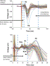
Overview of sciatic SSEP features and labeling of peaks. The top figure illustrates an overlay of all SSEP waveforms recorded over contralateral S1 hindlimb cortex in the DBS OFF state from the pre-stimulation baseline to 150 ms post stimulation. The graph labels stimulus artifact, short-latency range, and long-latency range. The bottom image shows a magnified view of the clustered waveforms from the short-latency range. Peaks were labeled an evaluated from P1A through N2 as shown.
Figure 4:
Figure 4:
Short- and long-latency RMS ratios summarized in a box and whisker plot. The ends of the box and whisker plots display the upper (75th) and lower percentiles (25th). The box spans the interquartile range (50th). The median is marked by a horizontal line within the interquartile range. The whiskers extend to the highest and lowest points observation. Circles outside of the box indicate outliers. Circles or positive signs within the boxes indicate mean. All analyzed data depicted was from channel 1 and 38 trials of 30 Hz and 27 trials of 100 Hz were included in analysis for both latencies. All RMS ratios were compared to a value of one and yielded insignificant (p value > 0.05, Wilcoxon) differences for both short and long latencies.
Figure 5:
Figure 5:
Peak to peak amplitude ratios summarized in box and whisker plots. The ends of the box and whisker plots display the upper (75th) and lower percentiles (25th). The box spans the interquartile range (50th). The median is marked by a horizontal line within the interquartile range. The whiskers extend to the highest and lowest points observation. Circles outside of the box indicate outliers. Circles or positive signs within the boxes indicate mean. Data analyzed from channel 1. 38 trials of 30 Hz and 27 trials of 100 Hz were included for analysis of individual peak ratios. Ratios of the peak-to-peak amplitudes for DBS ON to peak-to-peak amplitude of DBS OFF were calculated and compared to 1. 30 Hz trials resulted in one peak ratio of significance. P2-N1, denoted by the “#” symbol, was significantly greater than 1 (p = 0.044, GLMM). Remaining peak-to-peak ratios had no significance (p> 0.05, GLMM). 100 Hz trials resulted in two peaks of significance. P2-N1 (p = 0.018, GLMM), denoted by the “$” symbol, and P2-N2 (p = 0.003, GLMM), denoted by the “*” symbol, had ratios statistically significant from 1. Remaining peak- to-peak ratios had no significance (p> 0.05, GLMM).
Figure 6:
Figure 6:
Possible interactions between the ascending sensory and DTC pathways. Solid arrows depict better defined interactions and dashed lines depict controversial or non-confirmed interactions. Red arrows portray the main pathway of the sciatic nerve SSEP. Following electrical stimulation of the sciatic nerve, action potentials travel up the dorsal column of the spinal cord, synapse in nucleus gracilis, proceed to the thalamus, synapse in the VPLc nucleus, proceed to cortex, and synapse in primary somatosensory cortex. Other neural generators of cortical SSEPs have been suspected in S2, M1, and frontal lobe (not depicted). The spinocerebellar tract is also thought to contribute to SSEP transmission. DBS of the LCN activates cells which project to and synapse in multiple thalamic nuclei (notably MD, VLc, VPLo, and area X). The thalamic nuclei primarily project M1, but there is evidence for additional connections to pre-frontal, pre-motor, and posterior parietal cortex. Direct connections between the DTC pathway and S1 have been suspected but not explicitly demonstrated. Additional cortical-cortical synapses may be involved in DTC/ascending sensory communications. DTC activation has also been shown to send descending signals via the reticulospinal tract.

Similar articles

Cited by

References

    1. Yang Q, Tong X, Schieb L, Vaughan A, Gillespie C, Wiltz JL, King SC, Odom E, Merritt R, Hong Y, Vital signs: recent trends in stroke death rates—United States, 2000–2015, MMWR. Morbidity and mortality weekly report 66 (2017) 933. - PMC - PubMed
    1. Ward NS, Restoring brain function after stroke—bridging the gap between animals and humans, Nat Rev Neurol 13 (2017) 244–255. - PubMed
    1. Edwardson M, Lucas T, Carey J, Fetz E, New modalities of brain stimulation for stroke rehabilitation, Exp. Brain Res 224 (2013) 335–358. - PMC - PubMed
    1. Ting WK-C, Fadul FA-R, Fecteau S, Ethier C, Neurostimulation for Stroke Rehabilitation, Front. Neurosci 15 (2021) 583. - PMC - PubMed
    1. Hummel F, Cohen LG, Improvement of motor function with noninvasive cortical stimulation in a patient with chronic stroke, Neurorehabilitation and neural repair 19 (2005) 14–19. - PubMed

Publication types