. 2022 Jul 16;13(1):4137.
doi: 10.1038/s41467-022-31882-4.

Nanoparticle elasticity affects systemic circulation lifetime by modulating adsorption of apolipoprotein A-I in corona formation

Affiliations

Nanoparticle elasticity affects systemic circulation lifetime by modulating adsorption of apolipoprotein A-I in corona formation

Mingyang Li et al. Nat Commun. .

Abstract

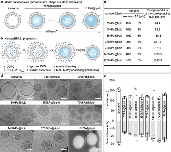
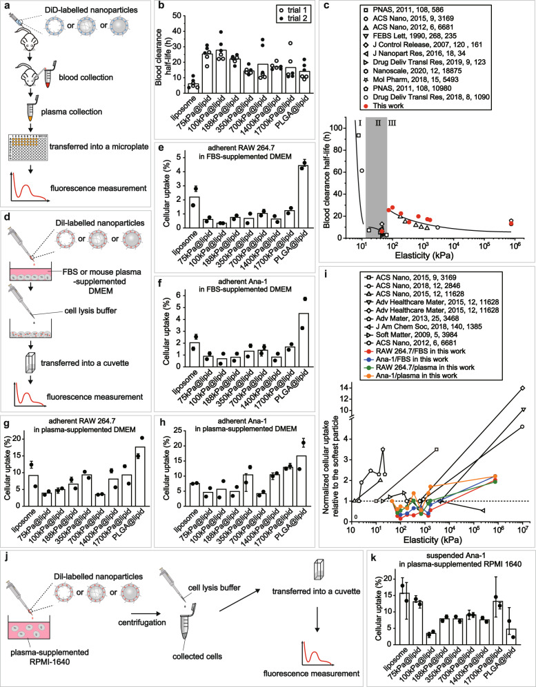
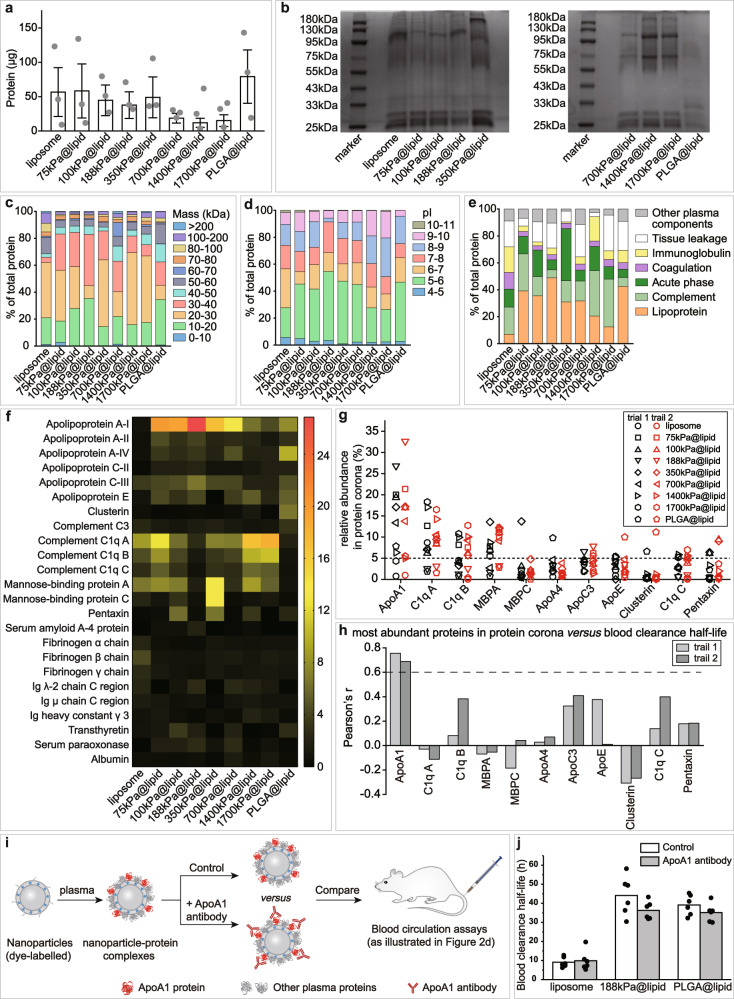
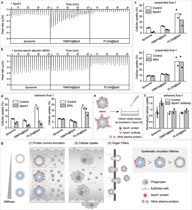
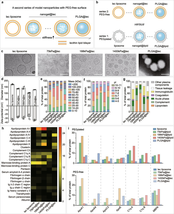
Nanoparticle elasticity is crucial in nanoparticles' physiological fate, but how this occurs is largely unknown. Using core-shell nanoparticles with a same PEGylated lipid bilayer shell yet cores differing in elasticity (45 kPa - 760 MPa) as models, we isolate the effects of nanoparticle elasticity from those of other physiochemical parameters and, using mouse models, observe a non-monotonic relationship of systemic circulation lifetime versus nanoparticle elasticity. Incubating our nanoparticles in mouse plasma provides protein coronas varying non-monotonically in composition depending on nanoparticle elasticity. Particularly, apolipoprotein A-I (ApoA1) is the only protein whose relative abundance in corona strongly correlates with our nanoparticles' blood clearance lifetime. Notably, similar results are observed when above nanoparticles' PEGylated lipid bilayer shell is changed to be non-PEGylated. This work unveils the mechanisms by which nanoparticle elasticity affects nanoparticles' physiological fate and suggests nanoparticle elasticity as a readily tunable parameter in future rational exploiting of protein corona.


Conflict of interest statement

The authors declare no competing interests.

Figures

Fig. 1
Fig. 1. Our PEGylated model nanoparticles.
a Schematic illustration on our model nanoparticles, which share a core-shell structure with a lipid bilayer shell of same composition while a core of tunable elasticity. b Schematic illustration on the preparation of a nanogel@lipid particle, whose elasticity can be tuned by adjusting its nanogel core’s crosslinking density. c Summary on the Young’s moduli of bulk gels which were prepared with corresponding hydrogel-precursor solutions used for preparing our nanogel@lipid particles. d Cryo-electron microscope (Cryo-EM) images of our liposome and nanogel@lipid nanoparticles, and transmission electron microscope (TEM) images of PLGA@lipid negatively stained with uranyl acetate. For each sample, microscopy images were taken in ≥5 different microscopy fields of view, and consistent results were observed. Scale bar = 50 nm. e Hydrodynamic diameters and surface zeta-potentials of our model nanoparticles. Bar heights are reported as average ± standard deviation (n = 3 independent experiments).
Fig. 2
Fig. 2. Effects of nanoparticle elasticity on blood circulation lifetime and cellular uptake.
a Schematic illustration on blood circulation test for obtaining blood retention profiles for our nanoparticles. b Blood clearance half-lives of our model nanoparticles. Bar heights are reported as averages of two independent trials (n = 3 biologically independent mice in each independent trial). c Plot on the relationship of blood clearance half-life versus nanoparticle elasticity, using results from this work and prior reports on this topic. d Schematic illustration on in vitro cellular uptake assays. Uptake efficiency of nanoparticles by murine macrophage e RAW264.7 and f Ana-1 cells. Bar heights are reported as averages of two independent trials, while data points are reported as average ± standard deviation (n = 3 in each independent trial). Uptake efficiency of our nanoparticles by adherent g RAW264.7 and h Ana-1 cells in mouse plasma-supplemented culture medium. Bar heights are reported as average of two independent trials, while data points are reported as average ± standard deviation (n = 3 in each independent trial). i Plots on the relationship of normalized cellular uptake relative to the softest particle in a same study versus nanoparticle elasticity, using results from this work and prior reports on this topic. j Schematic illustration on in vitro cellular uptake assays by suspended Ana-1 in mouse plasma-supplemented culture medium. k Uptake efficiency of our nanoparticles by suspended Ana-1 in mouse plasma-supplemented culture medium. Bar heights are reported as average of two independent trials, while data points are reported as average ± standard deviation (n = 3 in each independent trial).
Fig. 3
Fig. 3. Effects of nanoparticle elasticity on protein corona.
a Amounts of absorbed proteins on our nanoparticles (kept constant at 1 mg in lipid dose). Data points are reported as average ± standard deviation (n = 3 independent experiments). b Photographs of SDS-PAGE gels with absorbed proteins on our nanoparticles. The SDS-PAGE gel electrophoresis was only once. Classification of corona proteins according to c molecular weight, d calculated isoelectric point (pI), and e physiological functions. f Heat map of the most abundant proteins in protein coronas of our nanoparticles. g Distribution of the relative abundance in corona on different particles and h Pearson’s r between the relative abundance in corona and nanoparticle blood clearance half-life for proteins which exhibited a relative abundance of >5% on at least one nanoparticle. Pearson’s r of >0.6 and <−0.6 indicate strong positive and negative correlations, respectively. i Schematic illustration on blood circulation tests for examining the effects of pre-screening/pre-shielding the adsorbed ApoA1 in corona with ApoA1 antibody. j Blood clearance half-lives of 188 kPa@lipid with and without the adsorbed ApoA1 in corona pre-screened/pre-shielded by ApoA1 antibody, and those of liposome and PLGA@lipid in similar assays are included for reference. Bar heights are reported as average (n = 6 biologically independent mice).
Fig. 4
Fig. 4. Roles of Apolipoprotein A-I (ApoA1) in nanoparticle elasticity-dependence of systemic circulation lifetime.
Raw data from isothermal titration calorimetry (ITC) assays, in which a ApoA1 and b bovine serum albumin (BSA) solution was titrated into nanoparticle dispersions, using liposome, 188 kPa@lipid, and PLGA@lipid as representatives for nanoparticles with distinct elasticity. c Effects of the presence of ApoA1 and BSA (both at 50 μg/mL) on the uptake efficiency of our representative nanoparticles by suspended Ana-1 cells in RPMI-1640. Bar height are reported as average of two independent trials. d Effects of the presence of ApoA1 and BSA (both at 50 μg/mL) on the uptake efficiency of our representative nanoparticles by adherent Ana-1 cells in DMEM. Bar height are reported as average of two independent trials. e Schematic illustration on cellular uptake assays for examining the effects pre-screening/pre-shielding the adsorbed ApoA1 in corona with ApoA1 antibody. f Cellular uptake of a nanoparticle with and without ApoA1 in corona being pre-screened/pre-shielded with ApoA1 antibody, normalized relative to that of the particle without ApoA1 in corona being pre-screened/pre-shielded with ApoA1 antibody. Bar heights are reported as averages of two independent trials, while data points are reported as average ± standard deviation (n = 3 in each independent trial). g Schematic illustration on (1) ApoA1’s preferentiality in adsorbing onto nanoparticles of intermediate elasticity and consequently (2) suppressing their cellular uptake, which together with (3) softer nanoparticles’ higher tendency to pass organ filters lead to (right side) longer systemic circulation lifetime for nanoparticles of intermediate elasticity.
Fig. 5
Fig. 5. Nanoparticle elasticity overwhelms surface chemistry (PEG presence versus absence) in affecting protein corona.
Schematic illustrations on a our PEG-free model nanoparticles, which have a core-shell structure with a lipid bilayer shell composed of lecithin (lec) and a core of tunable elasticity, and b a comparison with PEGylated nanoparticles. c Cryo-EM images of our lec liposome and nanogel@lec particles and TEM image of our PLGA@lec. For each sample, microscopy images were taken in ≥5 different microscopy fields of view, and consistent results were observed. Scale bar = 100 nm. d Hydrodynamic diameter and surface and zeta-potentials of our PEG-free nanoparticles. Bar heights are reported as average ± standard deviation (n = 3 independent experiments). Classifications of corona proteins according to e molecular weight, f calculated isoelectric point (pI), and g physiological functions for our PEG-free nanoparticles. h Heat map of the most abundant proteins in the coronas on our PEG-free nanoparticles. i Comparison on the distribution of the relative abundance in corona on our (top) PEGylated and (bottom) PEG-free nanoparticles for proteins which exhibited a relative abundance in corona of >5% on at least one nanoparticle.

References

    1. Li H, Li Y, Jiao J, Hu H-M. Alpha-alumina nanoparticles induce efficient autophagy-dependent cross-presentation and potent antitumour response. Nat. Nanotechnol. 2011;6:645–650. doi: 10.1038/nnano.2011.153. - DOI - PMC - PubMed
    1. Moon JJ, et al. Interbilayer-crosslinked multilamellar vesicles as synthetic vaccines for potent humoral and cellular immune responses. Nat. Mater. 2011;10:243–251. doi: 10.1038/nmat2960. - DOI - PMC - PubMed
    1. Mi P, et al. A pH-activatable nanoparticle with signal-amplification capabilities for non-invasive imaging of tumour malignancy. Nat. Nanotechnol. 2016;11:724–730. doi: 10.1038/nnano.2016.72. - DOI - PubMed
    1. Peer D, et al. Nanocarriers as an emerging platform for cancer therapy. Nat. Nanotechnol. 2007;2:751–760. doi: 10.1038/nnano.2007.387. - DOI - PubMed
    1. Min Y, Caster JM, Eblan MJ, Wang AZ. Clinical translation of nanomedicine. Chem. Rev. 2015;115:11147–11190. doi: 10.1021/acs.chemrev.5b00116. - DOI - PMC - PubMed

Publication types