. 2022 Jul 16;12(1):12191.
doi: 10.1038/s41598-022-16253-9.

A human tissue-based assay identifies a novel carrion blowfly strain for maggot debridement therapy

Affiliations

A human tissue-based assay identifies a novel carrion blowfly strain for maggot debridement therapy

Takuma Yoshida et al. Sci Rep. .

Abstract

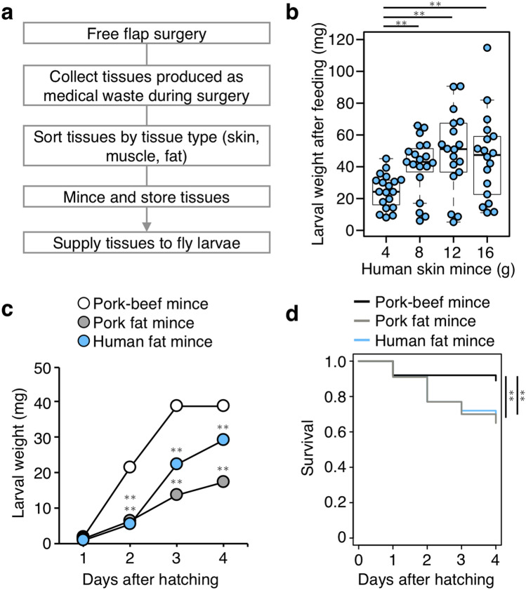
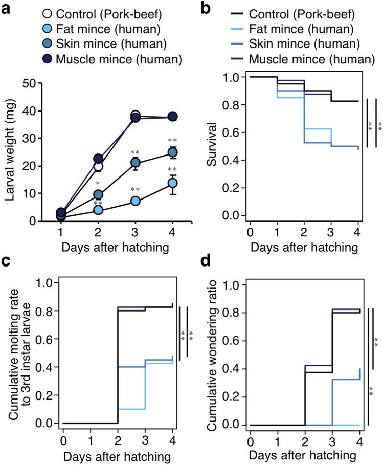
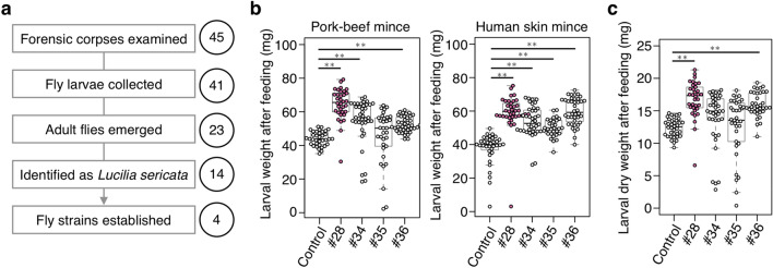
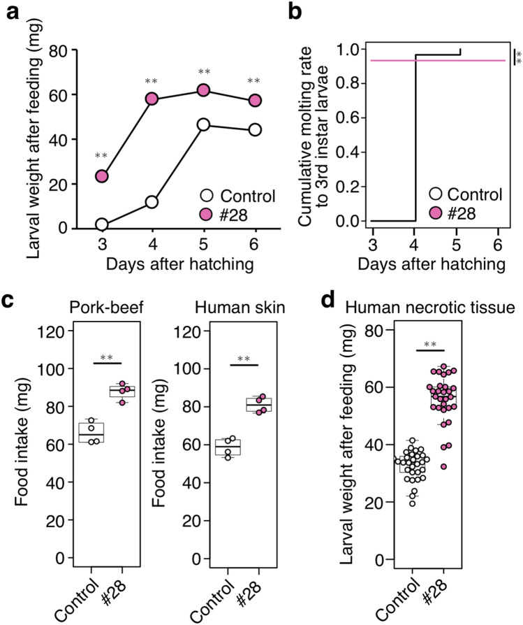
Maggot debridement therapy (MDT) is a form of therapeutic wound treatment in which live fly larvae are used intentionally to debride necrotic tissues. MDT has been widely used to treat chronic wounds in humans or animals, such as diabetic foot ulcers. Larvae of a carrion blowfly, Lucilia sericata (green bottle fly), debride wounds by consuming necrotic tissue and removing pathogenic bacteria, promoting effective wound healing. Most medical L. sericata strains were initially collected from natural environments using animal meat as bait and reared on artificial protein-rich media or ground meat. It remains to be examined which strain would be more appropriate for MDT, whereas any method for evaluating the fly's therapeutic potential in humans has not been available. A feeding assay was developed using minced human tissues obtained from surgical waste. To establish L. sericata strains highly eligible for MDT, carrion fly larvae were collected from 45 corpses subjected to forensic autopsy (such as decomposed bodies). Four corpse-derived L. sericata strains were obtained and evaluated using the feeding assay. One strain showed that its feeding activity was 1.4 times higher than the control strain used in conventional MDT. The body length of the adult fly of the corpse-derived strain was longer than the control, which was consistent with the observation that its cell size was enlarged. The human tissue-based assay developed in this study accurately evaluated the ability of fly larvae to debride necrotic wounds. The L. sericata strain newly established from human corpses harboring high feeding activity may offer a clinically significant improvement in MDT.


Conflict of interest statement

The authors declare no competing interests.

Figures

Figure 1
Figure 1
Development of a human tissue-based feeding assay for L. sericata larvae. (a) A flowchart describing human tissue preparation for making the diet of fly larvae. During free flap surgery, human tissues produced as medical waste were collected, sorted by type of tissue, minced, and supplied to L. sericata larvae. (b) Weight of larvae at 4 days after hatching and fed with human skin mince diet. The indicated amounts of diet were supplied to each group (n = 20). Each dot represents the weight of an individual larva. Boxplots: center line, median; box range, 25th–75th percentiles; whiskers denote minimum–maximum values. Student’s t-test, **p < 0.01. (c,d) Weight and survival ratio of larvae fed on 40 g of pork-beef mince, pork fat mince, or human fat mince. n = 100 per group. The mean values are shown ± SEM in (c). Student’s t-test (c), two-sided log-rank test (d). **p < 0.01. See also Supplementary Data 1 for data of experiment, exact sample sizes, and p values.
Figure 2
Figure 2
Human tissue-dependent growth and survivability of L. sericata larvae. (a,b) Weight and survival ratio of larvae fed on 16 g of pork-beef mince, human fat mince, human skin mince, or human muscle mince. n = 40 per group. The mean values are shown ± SEM in (a). Student’s t-test (a), two-sided log-rank test (b), *p < 0.05, **p < 0.01. (c,d) Developmental rates of larvae (third instar and wandering) fed on 16 g of pork-beef mince, human fat mince, human skin mince, or human muscle mince. n = 40 per group. Two-sided log-rank test, **p < 0.01. See also Supplementary Data 1 for data of experiment, exact sample sizes, and p values.
Figure 3
Figure 3
Analysis of carrion fly species collected from human corpses. (a) A flow chart of collecting fly larvae and identifying L. sericata from forensic corpses. Each number indicates the human corpse examined. Four L. sericata strains (#28, #34, #35, #36) were established. (b) Weight of larvae supplied with 24 g of pork-beef mince (left) or human skin mince (right). Weight was measured at 5 days (left) and 4 days (right) after hatching. n = 40 per group. (c) Dried weight of larvae at 5 days after hatching supplied with 24 g of pork-beef mince. n = 40 per group. In (b) and (c), each dot represents the weight of an individual larva. Boxplots: center line, median; box range, 25th–75th percentiles; whiskers denote minimum–maximum values. Student’s t-test, **p < 0.01. See also Supplementary Data 1 for data of experiment, exact sample sizes, and p values.
Figure 4
Figure 4
A corpse-derived fly strain that ingested human tissue effectively. (a,b) Weight (a) and developmental rate (third instar larvae) (b) of larvae supplied with 18 g of pork-beef mince. n = 40 per group. The mean values are shown ± SEM in (a). Student’s t-test (a), two-sided log-rank test (b). **p < 0.01. (c) Amount of food intake by larvae at 5 days after hatching supplied with 6 g of pork-beef mince (left) or human skin mince (right). n = 4 per group. (d) Weight of larvae at 5 days after hatching supplied with 18 g of human necrotic tissue mince. n = 30 per group. In (c) and (d), each dot represents the value of an individual larva. Boxplots: center line, median; box range, 25th–75th percentiles; whiskers denote minimum–maximum values. Student’s t-test, **p < 0.01. See also Supplementary Data 1 for data of experiment, exact sample sizes, and p values.
Figure 5
Figure 5
Correlation between feeding ability and body size of the corpse-derived flies. (a) Weight and length (major and minor axes) of pupa at 11 days after hatching. n = 30 per group. Bar = 4 mm. (b) Weight and length of adults at 26 days after hatching. n = 30 per group. Bar = 5 mm. (c) Trichome density and vein length of the wings of female adults at 26 days after hatching. n = 8 per group. (d) Size of cells in the fat body of larvae at 4 days after hatching. n = 40 per group. Magenta, phalloidin (actin); blue, TO-PRO-3 (nuclei). Bar = 100 µm. (e) Length of female adults of corpse-derived fly strains. Note that strain #34 was not included due to failure to maintain the strain. Each dot represents the value obtained from an individual specimen (ae). Boxplots: center line, median; box range, 25th–75th percentiles; whiskers denote minimum–maximum values. Student’s t-test, *p < 0.05, **p < 0.01. See also Supplementary Data 1 for data of experiment, exact sample sizes, and p values.
Figure 6
Figure 6
Cell proliferation activity of larval excretions and secretions (ES). (a) Proliferation rate of HFF cells treated with the indicated concentration of larval ES. n = 3 per group. (b) Dissolution rate of fibrin by culture supernatant of HFF cells treated with the indicated concentrations of larval ES. n = 3 per group. c Cell migration rate of HFF cells treated with the indicated concentration of larval ES. n = 3 per group. Each dot represents the value obtained from an individual specimen (ac). Each rate is expressed as the percentage of larval ES-treated condition versus PBS only treatment (shown as 100%). Boxplots: center line, median; box range, 25th–75th percentiles; whiskers denote minimum–maximum values. Student’s t-test, **p < 0.01. n.s., not significant. See also Supplementary Data 1 for data of experiment, exact sample sizes, and p values. (d) MA plots of RNA sequence reads of control strain, compared with strain #28. Genes with q-value < 0.05 are labeled in red. M, log2 fold change, A, averaged log2 reads.

Similar articles

References

    1. World Health Organization. Global report on diabetes. https://www.who.int/publications/i/item/9789241565257 (2016).
    1. Bhurosy T, Jeewon R. Overweight and obesity epidemic in developing countries: A problem with diet, physical activity, or socioeconomic status? ScientificWorldJournal. 2014;2014:964236. doi: 10.1155/2014/964236. - DOI - PMC - PubMed
    1. Misra A, Ramchandran A, Jayawardena R, Shrivastava U, Snehalatha C. Diabetes in South Asians. Diabet. Med. 2014;31(10):1153–1162. doi: 10.1111/dme.12540. - DOI - PubMed
    1. Boulton AJ, Vileikyte L, Ragnarson-Tennvall G, Apelqvist J. The global burden of diabetic foot disease. Lancet. 2005;366(9498):1719–1724. doi: 10.1016/S0140-6736(05)67698-2. - DOI - PubMed
    1. García-Morales E, Lázaro-Martínez JL, Martínez-Hernández D, Aragón-Sánchez J, Beneit-Montesinos JV, González-Jurado MA. Impact of diabetic foot related complications on the Health Related Quality of Life (HRQol) of patients—a regional study in Spain. Int. J. Low Extrem Wounds. 2011;10(1):6–11. doi: 10.1177/1534734611400257. - DOI - PubMed

Publication types