. 1987 May;131(2):165-74.
doi: 10.1002/jcp.1041310205.

Isolation of an autocrine growth factor from hepatoma HTC-SR cells

Isolation of an autocrine growth factor from hepatoma HTC-SR cells

P Ove et al. J Cell Physiol. 1987 May.

Abstract

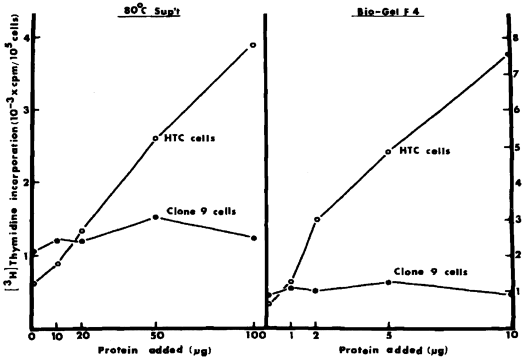
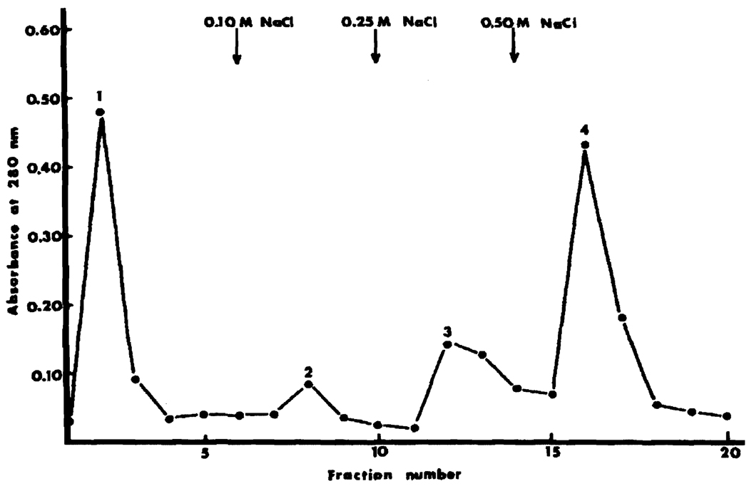
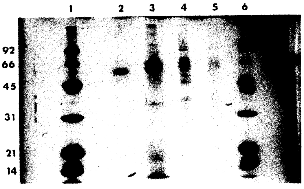
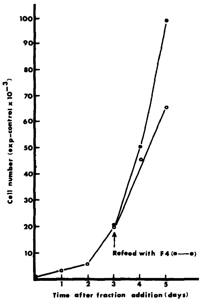
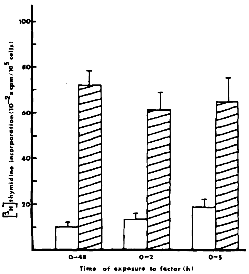
A growth factor has been isolated from HTC-SR rat hepatoma tissue culture cells which specifically stimulates DNA synthesis and cell proliferation of the HTC cells that produce it. The factor can be isolated from HTC cell conditioned medium or from an HTC cell extract. This autocrine factor has been purified 640-fold from a postmicrosomal supernatant by successive steps, involving ethanol precipitation, heating at 80 degrees C for 10 min, chromatography on a DEAE Bio-Gel A column, and chromatography on a heparin-sepharose affinity column. The major peak of activity eluted from the heparin column migrates as a single band on SDS-PAGE with an apparent Mr of 60,000. The factor is resistant to acid, heat, and neuraminidase but sensitive to trypsin, papain, and protease. The autocrine nature of the factor is indicated by the finding that several other types of cells do not respond with increased DNA synthesis. Mouse L-cells, BHK cells, Novikoff hepatoma cells, hepatocytes in primary culture, and an epithelial-like rat liver-derived cell line (Clone 9) were tested, and none of the cells could be stimulated. Small amounts of the factor could be extracted from the Clone 9 cells, however. This material had the same physical and purification properties as the factor extracted from HTC cells, but it did not stimulate DNA synthesis in Clone 9 cells, only in HTC cells. Addition of the factor resulted in an almost immediate stimulation of DNA synthesis in a proliferating HTC cell population. When the factor was added together with [3H]thymidine for 2 h, a significant stimulation of DNA synthesis was observed, provided the addition was made between 18 and 48 h after the cells had been plated. Autoradiographic studies indicated that the factor both accelerates DNA synthesis in cells already making DNA and increases the number of cells entering the S period. The stimulation of DNA synthesis was completely inhibited by 10 mM hydroxyurea, whether the factor was present for 2, 24, or 48 h in the culture. A significant increase in cell number due to addition of the factor was also observed. This accelerated proliferation was detectable only after the cells had been in culture for at least 48 h with the factor present.


Figures

Fig. 1
Fig. 1
Incorporation of [3H]thymidine at various times after factor addition. HTC cells were plated at a density of 5 × 10⁴ cells per 35-mm dish ± 50 µg/ml of 80°C supernatant, in complete medium supplemented with 10% FBS. For each time point, control dishes and dishes with factor were exposed for 2 h to 0.4 µCi [3H]thymidine per dish. Cells were harvested and [3H]thymidine incorporation was determined at indicated times. ○, Control dishes; ●, addition of 80°C supernatant. Each point represents the average of three determinations, with the SD indicated by the vertical bars.
Fig. 2
Fig. 2
Dose response of HTC and Clone 9 cells. Assay conditions were as described for Figure 1 but all dishes were exposed to 0.4 µCi [3H]thymidine from 46 to 48 h after plating and addition of growth factor. Both cell types were seeded at the same density of 5 × 10⁴ cells per dish. The numbers on the abscissa indicate the protein added per dish. Each point represents the average of at least three determinations, with individual values within 5% of the mean.
Fig. 3
Fig. 3
Elution profile of DEAE Bio-Gel A chromatography. The activity was recovered in fraction 4. Four milligrams protein of 80°C supernatant were applied to a 10 cm × 1 cm column equilibrated with 5 mM Tris – HCl, pH 7.6. Four milliliter fractions were collected. Following adsorption of the sample the column was washed with 20 ml 5 mM Tris – HCl, pH 7.6 and eluted stepwise with 20 ml each: 100 mM NaCl, 250 mM NaCl, and 500 mM NaCl each in 5 mM Tris, pH 7.6.
Fig. 4
Fig. 4
Elution profile of heparin-sepharose affinity chromatography. The activity eluted in fraction II. Three hundred micrograms DEAE Bio–Gel A fraction 4 were applied to a heparin-sepharose CL–6B column prepared as described in Materials and Methods. Following adsorption of the fraction the column was washed with 20 ml 5 mM Tris – HCl. pH 7.6 and the activity eluted with 100 mM NaCl: 5 mM res–HCl, pH 7.6. Two millimeter fractions were collected.
Fig. 5
Fig. 5
SDS polyacrylamide gel electrophoresis of the autocrine factor. Lanes 1 and 6, low Mr standards, lane 2, heparin-sepharose FII, 20 µg; lane 3, Bio-Gel F4, 50 µg; lane 4, 80°C supernatant, 30 µg; lane 5, 80 °C supernatant, 10 µg.
Fig. 6
Fig. 6
Stimulation of cell proliferation. HTC cells were subcultured and distributed at a cell density of 1.25 × 10⁴ cells per 35-mm dishes in complete medium ± 5 µg Bio-Gel fraction 4. No medium changes were made, and cells were dispersed by trypsin treatment at indicated times and counted with a Coulter counter. On day 3, some of the experimental dishes received an additional 5 µg factor. The values indicated were expressed as cell number in experimental dishes minus cell number in control dishes and were from duplicate determinations for each point.
Fig. 7
Fig. 7
DNA synthesis after short-term exposure to the factor. Assay conditions were as described in Figure 1. After 2 and 5 h, medium was removed from control and experimental dishes, the dishes were washed once, and complete medium without factor was added to both control and experimental dishes. Cells were exposed to [3H]thymidine from 46–48 h. The values indicated are the averages of five determinations with the SD. Open bar, control dishes; shaded bar, + 50 µg/ml 80°C supernatant.
Fig. 8
Fig. 8
Inhibition of DNA synthesis by hydroxyurea. Cells were plated as described in Figure 1. Five micrograms of Bio-Gel fraction 4 were added at indicated times. Hydroxyurea (10 mM final concentration) was added at 0 time to controls and at times indicated on the abscissa to the experimental dishes. All dishes received [3H]thymidine between 46–48 h. The values are the averages of five determinations with the SD. Shaded bar, − hydroxyurea; open bar, + hydroxyurea.

References

    1. Antoniades HN, Owen AJ. Growth factors and regulation of cell growth. Annu. Rev. Med. 1982;33:445–463. - PubMed
    1. Bowen-Pope DE, Vogel A, Ross R. Production of platelet-derived growth factor-like molecules and reduced expression of platelet-derived growth factor receptors accompany transformation by a wide spectrum of agents. Proc. Natl. Acad. Sci. USA. 1984;81:2396–2400. - PMC - PubMed
    1. Burk RR. A factor from a transformed cell line that affects migration. Proc. Natl. Acad. Sci. USA. 1973;70:369–372. - PMC - PubMed
    1. Childs CB, Proper JA, Tucker RF, Moses HL. Serum contains a platelet-derived transforming growth factor. Proc. Natl. Acad. Sci. USA. 1982;79:5312–5316. - PMC - PubMed
    1. Ciba Foundation Symposium 116. Growth Factors in Biology and Medicine. In: Evered D, Nugent J, Whelan J, editors. Pitman Publishing Ltd.; London. 1985. pp. 1–269. - PubMed

Publication types