. 2023 Aug;39(4):1509-1529.
doi: 10.1007/s10565-022-09741-1. Epub 2022 Jul 16.

Plk2-mediated phosphorylation and translocalization of Nrf2 activates anti-inflammation through p53/Plk2/p21cip1 signaling in acute kidney injury

Affiliations

Plk2-mediated phosphorylation and translocalization of Nrf2 activates anti-inflammation through p53/Plk2/p21cip1 signaling in acute kidney injury

Da-Eun Kim et al. Cell Biol Toxicol. 2023 Aug.

Abstract

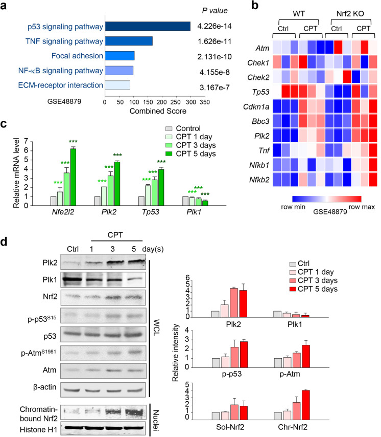
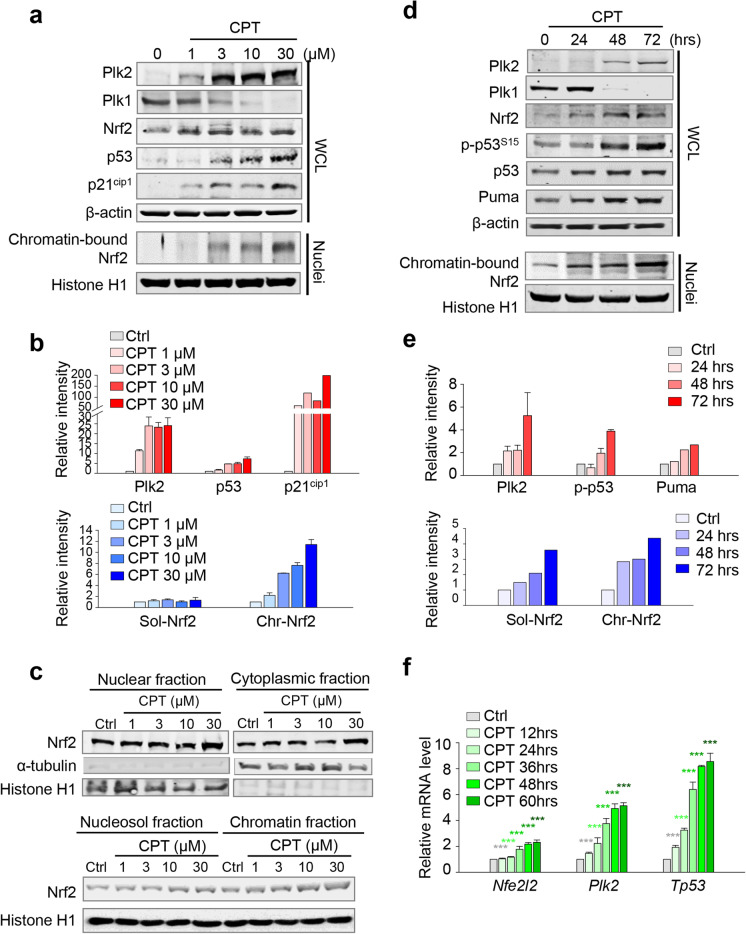
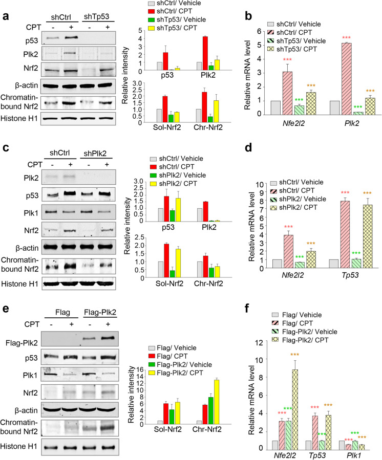
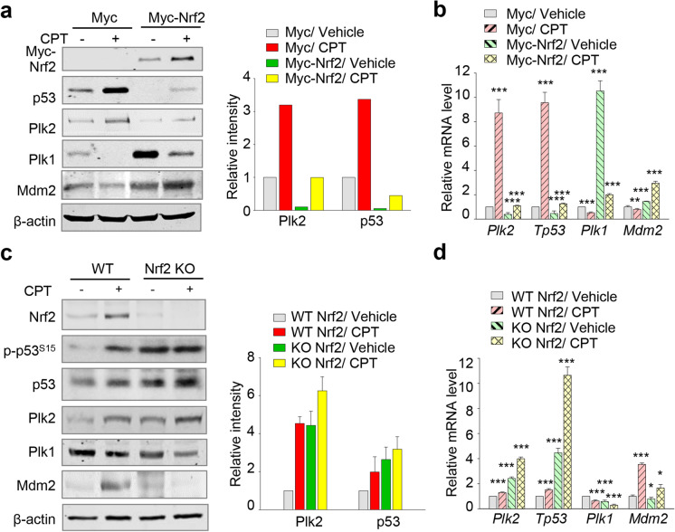
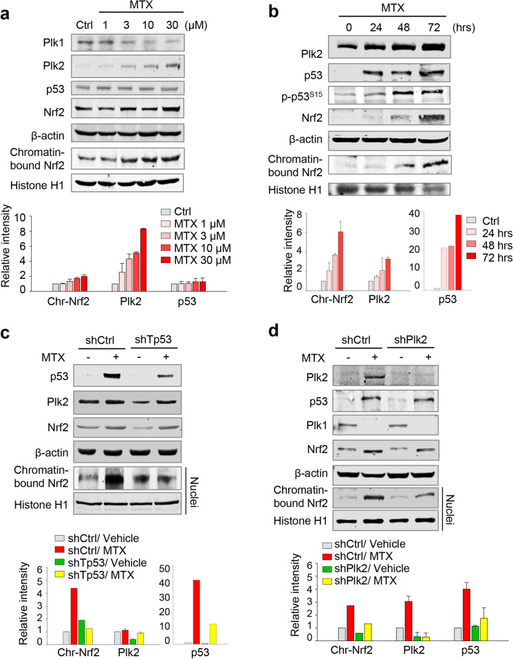
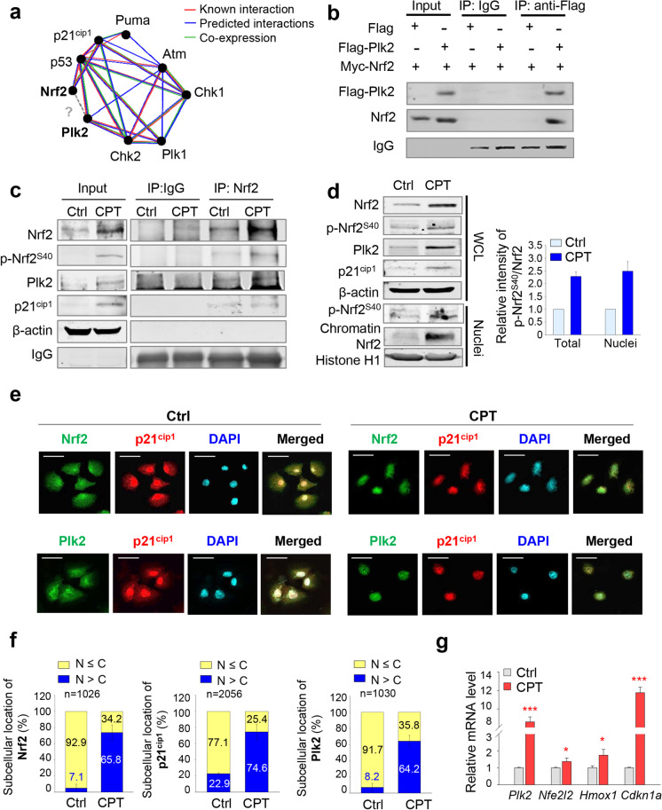
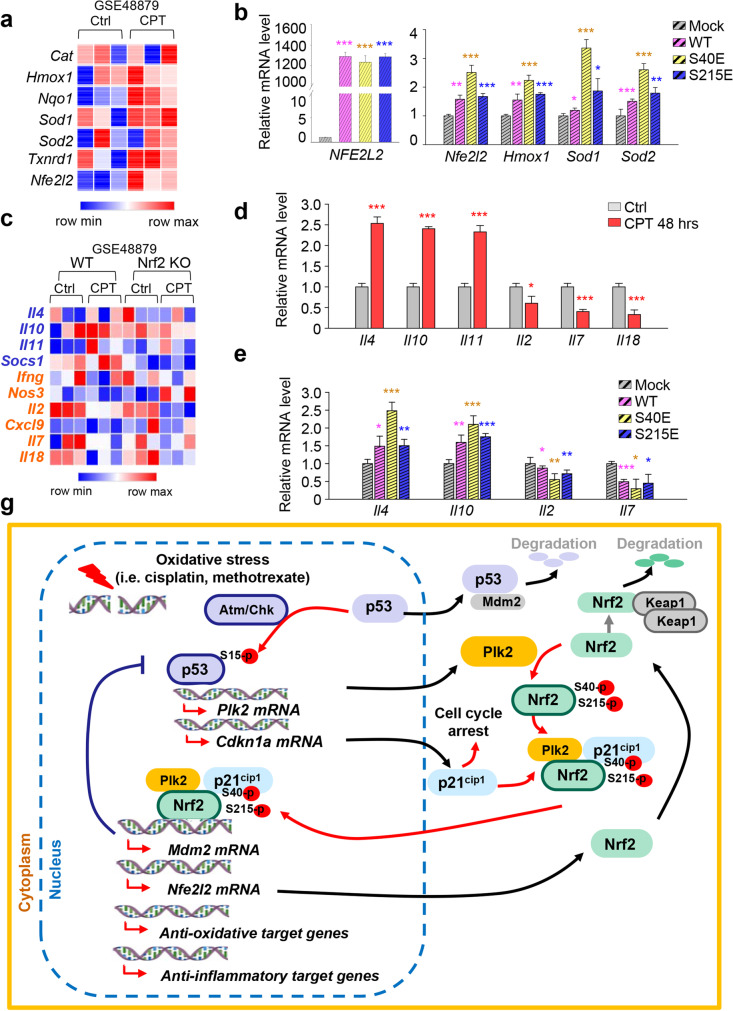
The Plk2 is a cellular stress-responsive factor that is induced in response to oxidative stress. However, the roles of Plk2 in acute kidney injury (AKI) have not been clarified. We previously found that Plk2 is an interacting factor of Nrf2 in response to cellular stress, since Plk2 is upregulated in the Nrf2-dependent network. Here, we show that the levels of p53, Plk2, p21cip1, and chromatin-bound Nrf2 were all upregulated in kidney tissues of mice or NRK52E cells treated with either cisplatin or methotrexate. Upregulation of Plk2 by p53 led to an increase of Nrf2 in both soluble and chromatin fractions in cisplatin-treated NRK52E cells. Consistently, depletion of Plk2 suppressed the levels of Nrf2. Of note, Plk2 directly phosphorylated Nrf2 at Ser40, which facilitated its interaction with p21cip1 and translocation into the nuclei for the activation of anti-oxidative and anti-inflammatory factors in response to AKI. Together, these findings suggest that Plk2 may serve as an anti-oxidative and anti-inflammatory regulator through the phosphorylation and activation of Nrf2 to protect kidney cells from kidney toxicants and that Plk2 and Nrf2 therefore work cooperatively for the protection and survival of kidney cells from harmful stresses.

Keywords: Acute kidney injury; Anti-inflammation; Nrf2; Plk2; p53.


Conflict of interest statement

The authors declare no competing interests.

The authors declare no competing interests.

Figures

Fig. 1
Fig. 1
Cisplatin-induced AKI increases the expressions of Plk2, p53, and Nrf2, which upregulate the levels of nuclear chromatin-bound Nrf2 in mouse kidney. a The previously published microarray dataset (GSE48879) was analyzed by KEGG pathway analysis. Analysis of transcriptome data for gene probes with fold changes of more than 1.5 (general cutoff point for DEGs) and p values are displayed. b A heatmap analysis was performed for Atm, Chek1, Chek2, Tp53, Cdkn1a, Bbc3, Plk2, Tnf, Nfkb1, and Nfkb2 using a published transcriptome dataset (GSE48879) from cisplatin (CPT)–treated mice. cd Kidney samples of the mice were taken at 1, 3, and 5 day(s) after a single injection of cisplatin (15 mg/kg, i.p., n = 4 or 5 per group). c Quantitative RT (qRT)-PCR was performed to determine the mRNA levels of Plk1, Plk2, Tp53, and Nfe2l2 in the kidney of mice treated with cisplatin for 1, 3, and 5 day(s). The relative expression of each gene was plotted. ***p < 0.001. d Immunoblot analysis was performed using the lysates of kidney cortex of the mice injected with cisplatin for 1, 3, and 5 day(s). The expressions of Plk2, Plk1, Nrf2, p-p53 (Ser15), p53, p-Atm (Ser1981), Atm, Histone H1, and β-actin were measured by immunoblottings. Chromatin fractions were isolated from whole-cell lysates, and the expression of Nrf2 protein was analyzed by immunoblot. Histone H1 was used as a loading control for the nuclear fraction
Fig. 2
Fig. 2
Cisplatin treatment increases levels of Plk2, p53, and Nrf2 in rat kidney NRK52E cells. ac Cisplatin (CPT) was treated at various concentrations of NRK52E rat renal proximal tubular cells at the indicated concentrations. a Immunoblot was performed with whole lysates using antibodies against Plk1, Plk2, p53, p21cip1, Nrf2, Histone H1, and β-actin. For detection of chromatin-bound proteins, Histone H1 was used as a loading control. b The relative band intensity values of Plk2, p53, p21cip1, soluble Nrf2, and chromatin-bound Nrf2 were measured using LI-COR Odyssey software. c A fractionation assay was performed. Nuclear and cytoplasmic fractions were prepared from whole cells; nuclear soluble fraction and chromatin fraction were from the nuclear fraction. Nrf2 protein was analyzed by immunoblot. Histone H1 was used as a loading control of the nuclear fraction. de Cisplatin was treated to NRK52E cells at the indicated times. d Immunoblot was performed with whole lysate using antibodies against Plk2, Plk1, p-p53 (S15), p53, Puma, Nrf2, β-actin, and Histone H1. For detection of chromatin-bound proteins, Histone H1 was used as a loading control. e The relative band intensity values of Plk2, p-p53, Puma, soluble Nrf2, and chromatin-bound Nrf2 were analyzed using LI-COR Odyssey software. f qRT-PCR assays were performed using primers for Plk2, Tp53, and Nfe2l2. ***p < 0.001 compared with control
Fig. 3
Fig. 3
Regulation of Plk2 by p53 results in the activation of Nrf2 in response to cisplatin-treated NRK52E cells. ab NRK52E cells were infected with lentiviral shRNA targeting rat Tp53. Twenty-four hours after infection, the cells were treated with/without 30 μM cisplatin for 48 h. a Immunoblot analysis was performed with antibodies for p53, Plk2, Nrf2, β-actin, and Histone H1. Histone H1 was used as a loading control for the chromatin fraction. The relative band intensity values of p53, Plk2, soluble Nrf2, and chromatin-bound Nrf2 were measured using LI-COR Odyssey software. b mRNA was prepared from NRK52E cell lysates for qRT-PCR analysis. The mRNA expression levels of Plk2 and Nfe2l2 were determined. ***p < 0.001. cd Rat Plk2 shRNA was introduced in NRK52E cells. Twenty-four hours after infection, the cells were treated with/without 30 μM cisplatin for 48 h. c Immunoblot analysis was performed with antibodies for Plk2, p53, Plk1, Nrf2, β-actin, and Histone H1. The relative band intensity values of p53, Plk2, soluble Nrf2, and chromatin-bound Nrf2 were measured. d mRNA was prepared from NRK52E cell lysates for qRT-PCR analysis. The mRNA expression levels of Nfe2l2 and Tp53 were determined and plotted. ***p < 0.001 compared with vehicle cells treated with control shRNA (shCtrl). ef Flag-tagged Plk2 proteins were expressed in NRK52E cells. Twenty-four hours after transfection, 30 μM cisplatin was treated to the cells for 48 h. e Immunoblot analysis was performed with antibodies for Flag, p53, Plk1, Nrf2, β-actin, and Histone H1. The relative band intensity values of soluble Nrf2 and chromatin-bound Nrf2 were analyzed using LI-COR Odyssey software. f qRT-PCR was performed to evaluate the mRNA levels of rat Nfe2l2, Tp53, and Plk1 in NRK52E cells. ***p < 0.001 compared with vehicle cells expressing Flag-mock control
Fig. 4
Fig. 4
Nrf2 negatively regulates the expression of p53 and Plk2 by upregulating Mdm2. ab Myc-tagged Nrf2 proteins were expressed in NRK52E cells. Twenty-four hours after transfection, the cells were treated with/without 30 μM cisplatin for 48 h. a Immunoblot analysis was performed with antibodies for Myc, p53, Plk2, Plk1, Mdm2, and β-actin. The relative band intensity values of p53 and Plk2 were analyzed using LI-COR Odyssey software. b qRT-PCR was performed to evaluate the mRNA levels of rat Plk2, Tp53, Plk1, and Mdm2 in NRK52E cells. ***p < 0.001. cd The kidney cortex tissues of Nrf2-/- mice were taken 3 days after a single injection of cisplatin (15 mg/kg, i.p., n = 4 or 5 per group). c Using the lysates of kidney cortex of mice, immunoblot analysis was performed. The expressions of Nrf2, p-p53 (Ser15), p53, Plk1, Plk2, Mdm2, and β-actin were measured using LI-COR Odyssey software and plotted. d qRT-PCR was performed to determine the mRNA levels of Plk2, Tp53, Plk1, and Mdm2 in the kidney of mice treated with cisplatin. The relative expression of each gene was plotted. ***p < 0.001 compared with the vehicle of Nrf2 WT mice samples
Fig. 5
Fig. 5
Upregulation of Plk2 and chromatin-bound Nrf2 in methotrexate-treated NRK52E cells. a Methotrexate (MTX) was treated at the indicated concentrations in NRK52E cells. Immunoblot was performed with whole lysate using specific antibodies for Plk1, Plk2, p53, Nrf2, Histone H1, and β-actin. The relative band intensity values were quantified and plotted. b NRK52E cells were treated with 30-μM methotrexate for the indicated times. Immunoblot analysis was performed for p53, p-p53 (Ser15), Plk2, Nrf2, β-actin, and Histone H1. Chromatin-bound Nrf2 and Histone H1 were detected using nuclear fractions. The relative band intensity values were quantified and plotted. c NRK52E cells were infected with lentivirus expressing rat Tp53 shRNA. Twenty-four hours after infection, the cells were treated with/without 30 μM methotrexate for 48 h. Immunoblot analysis was performed with antibodies for p53, Plk2, Nrf2, β-actin, and Histone H1. The relative band intensity values of p53, Plk2, and chromatin-bound Nrf2 were quantified and plotted. d Twenty-four hours after infection with lentiviral Plk2 shRNA in NRK52E cells, the cells were treated with 30 μM methotrexate for 48 h. Immunoblot analysis was performed with antibodies for Plk2, p53, Plk1, Nrf2, Histone H1, and β-actin. The relative band intensity values were plotted
Fig. 6
Fig. 6
Phosphorylated Nrf2 at Ser40 by Plk2 interacts with p21cip1, which accelerates translocalization into the nuclei in cisplatin-induced stress of NRK52E cells. a The network of p53 signaling factors was extracted from the GeneMANIA database. Red line, known interaction; blue line, predicted interaction; and green line, co-expression. b Flag-tagged Plk2 and/or Myc-tagged Nrf2 were expressed in HEK293 cells. Immunoprecipitation was performed with anti-Flag antibody. Immunoblot was performed with anti-Plk2 and anti-Nrf2 antibodies. c NRK52E cells were grown for 48 h in the presence of 30-μM cisplatin. Immunoprecipitation was performed with anti-Nrf2 antibody, and immunoblot was performed using specific antibodies for Plk2, Nrf2, p-Nrf2 (S40), p21cip1, and β-actin. d Cisplatin at 30 μM was treated to NRK52E cells for 48 h. Immunoblot was performed with whole lysates or nuclear fractions using specific antibodies for Plk2, Nrf2, p-Nrf2 (S40), p21cip1, β-actin, and Histone H1. e Cells were grown for 48 h in the presence or absence of 30-μM cisplatin. The cells were fixed with 4% paraformaldehyde and stained for Nrf2 or Plk2 (green), p21cip1 (red), and DAPI (blue). Bars, 25 μM. f The population of cells showing the indicated subcellular location of Nrf2, Plk2, and p21.cip1 of Fig. 6e was quantified in the presence or absence of 30-μM cisplatin. g qRT-PCR was performed using NRK52E cells in the presence or absence of 30-μM cisplatin. Relative mRNA expression levels of Plk2, Nfe2l2, Hmox1, and Cdkn1a were determined and plotted. *p < 0.05; ***p < 0.001 compared with control cells
Fig. 7
Fig. 7
Plk2-dependent phosphorylation of Nrf2 facilitates the expression of anti-oxidative and anti-inflammatory genes in rat kidney NRK52E cells. a A heatmap analysis was performed for Cat, Hmox1, Nqo1, Sod1, Sod2, Txnrd1, and Nfe2l2 using a published transcriptome dataset (GSE48879) from cisplatin-treated mice. b qRT-PCR was performed for human NFE2L2, rat Nfe2l2, Hmox1, Sod1, and Sod2 in NRK52E cells expressing Myc-tagged mock, wild-type (WT) human Nrf2, S40E, or S215E mutant. NFE2L2 is for the exogenous Myc-tagged human Nrf2. *p < 0.05; **p < 0.01; ***p < 0.001 c A heatmap analysis was performed for Il4, Il10, Il11, Il2, Il7, Il18, Socs1, Ifng, Nos3, and Cxcl9 using a published transcriptome dataset (GSE48879) from cisplatin (CPT)–treated wild-type (WT) and Nrf2 KO mice. d qRT-PCR was performed for Il4, Il10, Il11, Il2, Il7, and Il18 in NRK52E cells treated with 30-μM cisplatin for 48 h. *p < 0.05; ***p < 0.001. e qRT-PCR was performed for Il4, Il10, Il2, and Il7 in NRK52E cells expressing mock, wild type (WT), S40E, and S215E of Nrf2. *p < 0.05; **p < 0.01; ***p < 0.001 compared with control cells expressing myc-mock. f A plausible model of Nrf2 activation signaling: p53-mediated upregulation of Plk2 and p21cip1 induces the activation of Nrf2 through phosphorylation and recruitment to the nuclei for the expression of target genes related to anti-oxidative and anti-inflammatory signaling

Similar articles

Cited by

References

    1. Basu BA, Woolard MD, Johnson CL. Involvement of protein kinase C-delta in DNA damage-induced apoptosis. Cell Death Differ. 2001;8:899–908. doi: 10.1038/sj.cdd.4400885. - DOI - PubMed
    1. Benetatos L, Dasoula A, Hatzimichael E, Syed N, Voukelatou M, Dranitsaris G, Bourantas KL, Crook T. Polo-like kinase 2 (SNK/PLK2) is a novel epigenetically regulated gene in acute myeloid leukemia and myelodysplastic syndromes: genetic and epigenetic interactions. Ann Hematol. 2011;90:1037–1045. doi: 10.1007/s00277-011-1193-4. - DOI - PubMed
    1. Boyanapalli SS, Paredes-Gonzalez X, Fuentes F, Zhang C, Guo Y, Pung D, Saw CL, Kong AN. Nrf2 knockout attenuates the anti-inflammatory effects of phenethyl isothiocyanate and curcumin. Chem Res Toxicol. 2014;27:2036–2043. doi: 10.1021/tx500234h. - DOI - PMC - PubMed
    1. Burns TF, Fei P, Scata KA, Dicker DT, El-Deiry WS. Silencing of the novel p53 target gene Snk/Plk2 leads to mitotic catastrophe in paclitaxel (taxol)-exposed cells. Mol Cell Biol. 2003;23:5556–5571. doi: 10.1128/MCB.23.16.5556-5571.2003. - DOI - PMC - PubMed
    1. Cha HJ, Yim H. The accumulation of DNA repair defects is the molecular origin of carcinogenesis. Tumour Biol. 2013;34:3293–3302. doi: 10.1007/s13277-013-1038-y. - DOI - PubMed

Publication types