

Vitamin C attenuates predisposition to high-fat diet-induced metabolic dysregulation in GLUT10-deficient mouse model
- PMID: 35842612
- PMCID: PMC9288715
- DOI: 10.1186/s12263-022-00713-y
Vitamin C attenuates predisposition to high-fat diet-induced metabolic dysregulation in GLUT10-deficient mouse model
Abstract
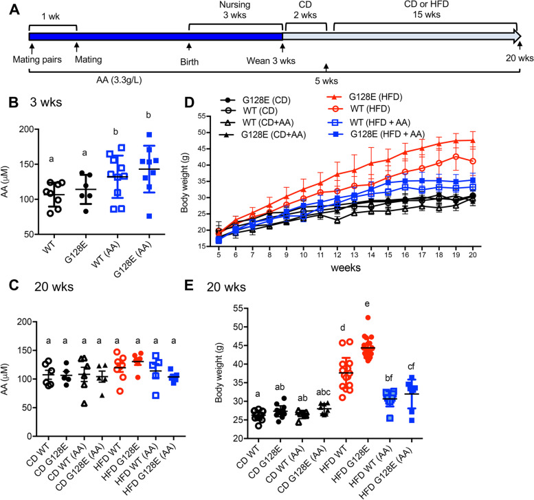
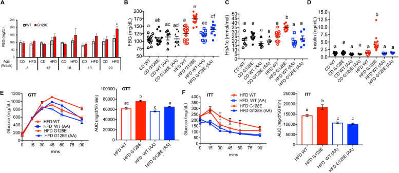
Background: The development of type 2 diabetes mellitus (T2DM) is highly influenced by complex interactions between genetic and environmental (dietary and lifestyle) factors. While vitamin C (ascorbic acid, AA) has been suggested as a complementary nutritional treatment for T2DM, evidence for the significance and beneficial effects of AA in T2DM is thus far inconclusive. We suspect that clinical studies on the topic might need to account for combination of genetic and dietary factors that could influence AA effects on metabolism. In this study, we tested this general idea using a mouse model with genetic predisposition to diet-induced metabolic dysfunction. In particular, we utilized mice carrying a human orthologous GLUT10G128E variant (GLUT10G128E mice), which are highly sensitive to high-fat diet (HFD)-induced metabolic dysregulation. The genetic variant has high relevance to human populations, as genetic polymorphisms in glucose transporter 10 (GLUT10) are associated with a T2DM intermediate phenotype in nondiabetic population.
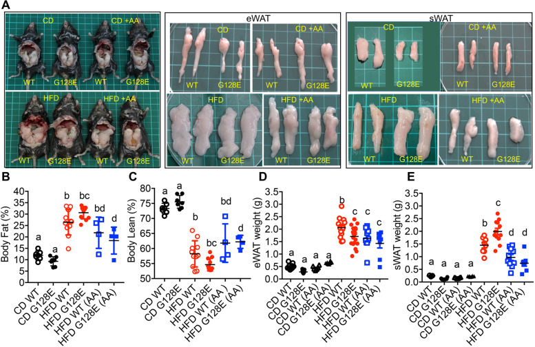
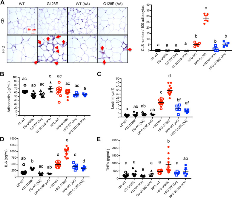
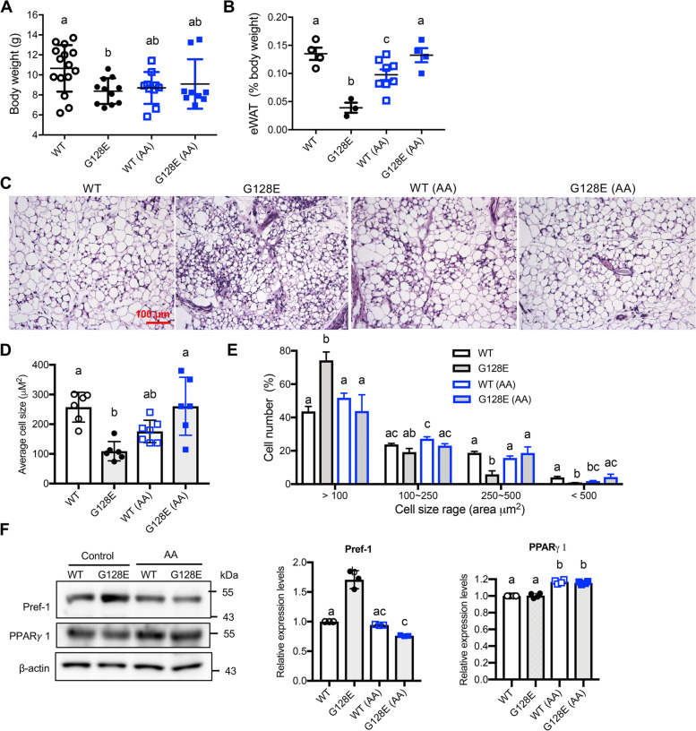
Results: We investigated the impacts of AA supplementation on metabolism in wild-type (WT) mice and GLUT10G128E mice fed with a normal diet or HFD. Overall, the beneficial effects of AA on metabolism were greater in HFD-fed GLUT10G128E mice than in HFD-fed WT mice. At early postnatal stages, AA improved the development of compromised epididymal white adipose tissue (eWAT) in GLUT10G128E mice. In adult animals, AA supplementation attenuated the predisposition of GLUT10G128E mice to HFD-triggered eWAT inflammation, adipokine dysregulation, ectopic fatty acid accumulation, metabolic dysregulation, and body weight gain, as compared with WT mice.
Conclusions: Taken together, our findings suggest that AA has greater beneficial effects on metabolism in HFD-fed GLUT10G128E mice than HFD-fed WT mice. As such, AA plays an important role in supporting eWAT development and attenuating HFD-induced metabolic dysregulation in GLUT10G128E mice. Our results suggest that proper WAT development is essential for metabolic regulation later in life. Furthermore, when considering the usage of AA as a complementary nutrition for prevention and treatment of T2DM, individual differences in genetics and dietary patterns should be taken into account.
Keywords: Genetic predisposition; High-fat diet; Type 2 diabetes mellitus; Vitamin C; White adipose tissue.
© 2022. The Author(s).
Conflict of interest statement
The authors declare that they have no competing interests.
Figures


Similar articles
-
Glucose transporter 10 modulates adipogenesis via an ascorbic acid-mediated pathway to protect mice against diet-induced metabolic dysregulation.PLoS Genet. 2020 May 26;16(5):e1008823. doi: 10.1371/journal.pgen.1008823. eCollection 2020 May. PLoS Genet. 2020. PMID: 32453789 Free PMC article.
-
Untargeted metabolomics uncovers metabolic dysregulation and tissue sensitivity in ACE2 knockout mice.Heliyon. 2024 Mar 8;10(6):e27472. doi: 10.1016/j.heliyon.2024.e27472. eCollection 2024 Mar 30. Heliyon. 2024. PMID: 38496880 Free PMC article.
-
Dual specificity phosphatase 6 deficiency is associated with impaired systemic glucose tolerance and reversible weight retardation in mice.PLoS One. 2017 Sep 5;12(9):e0183488. doi: 10.1371/journal.pone.0183488. eCollection 2017. PLoS One. 2017. PMID: 28873424 Free PMC article.
-
Ascorbic acid inhibits visceral obesity and nonalcoholic fatty liver disease by activating peroxisome proliferator-activated receptor α in high-fat-diet-fed C57BL/6J mice.Int J Obes (Lond). 2019 Aug;43(8):1620-1630. doi: 10.1038/s41366-018-0212-0. Epub 2018 Oct 3. Int J Obes (Lond). 2019. PMID: 30283077
-
P2Y2 Receptor Promotes High-Fat Diet-Induced Obesity.Front Endocrinol (Lausanne). 2020 Jun 3;11:341. doi: 10.3389/fendo.2020.00341. eCollection 2020. Front Endocrinol (Lausanne). 2020. PMID: 32582029 Free PMC article.
Cited by
-
The Role of Natural Antioxidant Products That Optimize Redox Status in the Prevention and Management of Type 2 Diabetes.Antioxidants (Basel). 2023 May 23;12(6):1139. doi: 10.3390/antiox12061139. Antioxidants (Basel). 2023. PMID: 37371869 Free PMC article. Review.
-
Progesterone levels associated with proteinuria in male diabetes mellitus patients: A cross-sectional retrospective study.J Diabetes Investig. 2023 May;14(5):669-674. doi: 10.1111/jdi.13992. Epub 2023 Feb 23. J Diabetes Investig. 2023. PMID: 36824009 Free PMC article.
-
Transcriptomic and metabonomic profiling unveils the mechanism of Tartary buckwheat and kiwi co-fermentation products in hyperlipidemia treatment.Front Pharmacol. 2025 May 30;16:1572593. doi: 10.3389/fphar.2025.1572593. eCollection 2025. Front Pharmacol. 2025. PMID: 40520184 Free PMC article.
-
Plant-based and sustainable diet: A systematic review of its impact on obesity.Obes Rev. 2025 Jun;26(6):e13901. doi: 10.1111/obr.13901. Epub 2025 Jan 29. Obes Rev. 2025. PMID: 39888238 Free PMC article.
References
-
- McAllister K, Mechanic LE, Amos C, Aschard H, Blair IA, Chatterjee N, Conti D, Gauderman WJ, Hsu L, Hutter CM, et al. Current challenges and new opportunities for gene-environment interaction studies of complex diseases. Am J Epidemiol. 2017;186(7):753–761. doi: 10.1093/aje/kwx227. - DOI - PMC - PubMed
Grants and funding
LinkOut - more resources
Full Text Sources
Research Materials
