. 2022 Jul 16;13(1):318.
doi: 10.1186/s13287-022-02999-6.

Notch-activated mesenchymal stromal/stem cells enhance the protective effect against acetaminophen-induced acute liver injury by activating AMPK/SIRT1 pathway

Affiliations

Notch-activated mesenchymal stromal/stem cells enhance the protective effect against acetaminophen-induced acute liver injury by activating AMPK/SIRT1 pathway

Mengxue Yu et al. Stem Cell Res Ther. .

Abstract

Background: Notch signaling plays important roles in regulating innate immunity. However, little is known about the role of Notch in mesenchymal stromal/stem cell (MSC)-mediated immunomodulation during liver inflammatory response.

Methods: Notch activation in human umbilical cord-derived MSCs was performed by a tissue culture plate coated with Notch ligand, recombinant human Jagged1 (JAG1). Mice were given intravenous injection of Notch-activated MSCs after acetaminophen (APAP)-induced acute liver injury. Liver tissues were collected and analyzed by histology and immunohistochemistry.

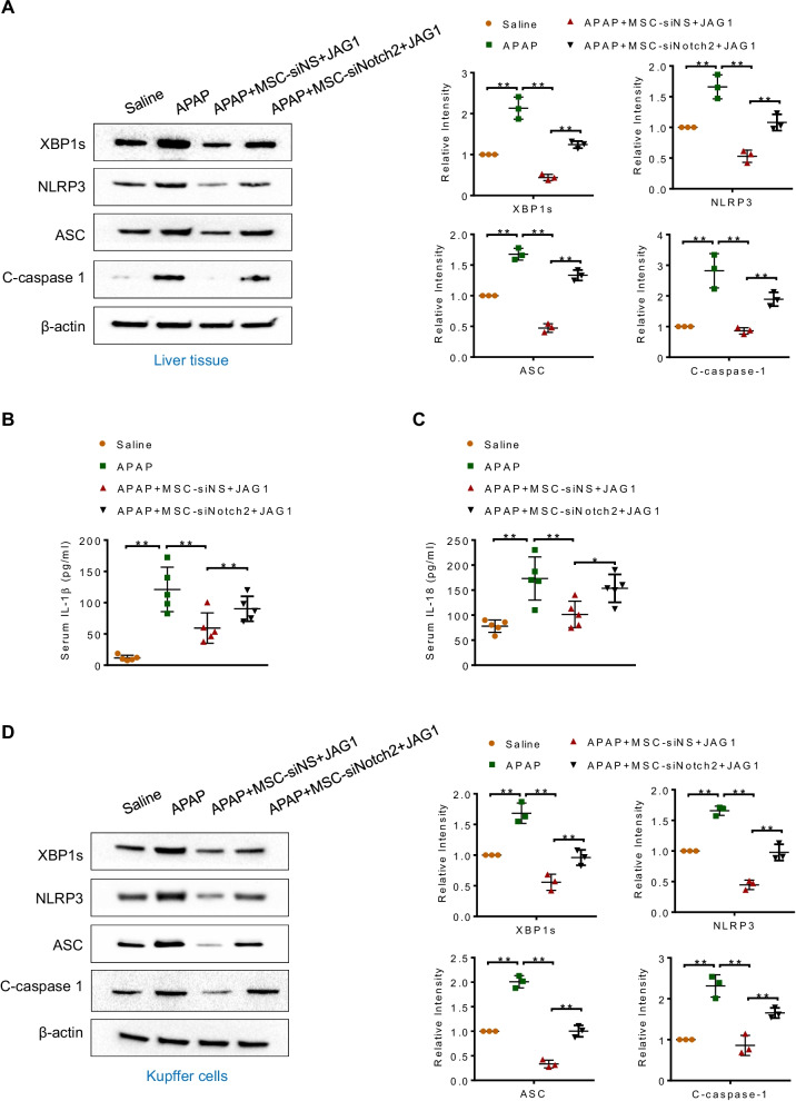
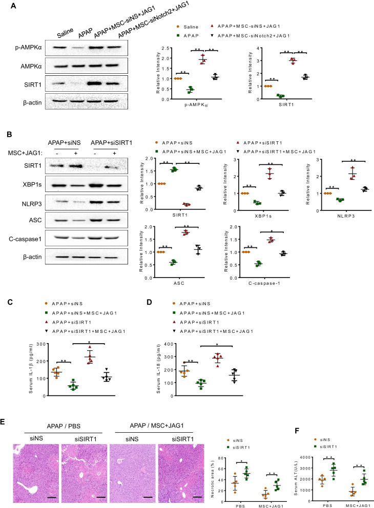
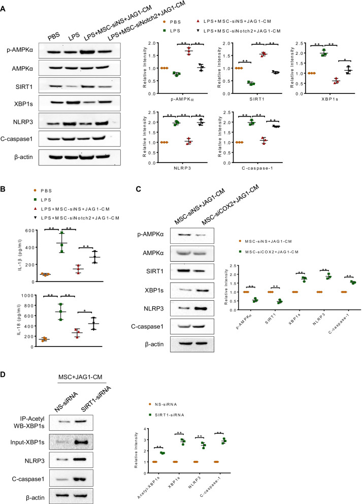
Results: MSC administration reduced APAP-induced hepatocellular damage, as manifested by decreased serum ALT levels, intrahepatic macrophage/neutrophil infiltration, hepatocellular apoptosis and proinflammatory mediators. The anti-inflammatory activity and therapeutic effects of MSCs were greatly enhanced by Notch activation via its ligand JAG1. However, Notch2 disruption in MSCs markedly diminished the protective effect of MSCs against APAP-induced acute liver injury, even in the presence of JAG1 pretreatment. Strikingly, Notch-activated MSCs promoted AMP-activated protein kinase (AMPKα) phosphorylation, increased the sirtuins 1 (SIRT1) deacetylase expression, but downregulated spliced X-box-binding protein 1 (XBP1s) expression and consequently reduced NLR family pyrin domain-containing 3 (NLRP3) inflammasome activation. Furthermore, SIRT1 disruption or XBP1s overexpression in macrophages exacerbated APAP-triggered liver inflammation and augmented NLRP3/caspase-1 activity in MSC-administrated mice. Mechanistic studies further demonstrated that JAG1-pretreated MSCs activated Notch2/COX2/PGE2 signaling, which in turn induced macrophage AMPK/SIRT1 activation, leading to XBP1s deacetylation and inhibition of NLRP3 activity.

Conclusions: Activation of Notch2 is required for the ability of MSCs to reduce the severity of APAP-induced liver damage in mice. Our findings underscore a novel molecular insights into MSCs-mediated immunomodulation by activating Notch2/COX2/AMPK/SIRT1 pathway and thus provide a new strategy for the treatment of liver inflammatory diseases.

Keywords: Acute liver injury; Mesenchymal stromal/stem cell; Notch signaling; SIRT1; XBP1.


Conflict of interest statement

The authors declare that they have no competing interest.

Figures

Fig. 1
Fig. 1
Notch-activated MSCs enhance the protective effects against APAP-induced hepatotoxicity. A Mice were subjected to APAP-challenged liver injury. Some mice were injected via the tail vein with human umbilical cord-derived MSCs, fibroblasts (1 × 106) or PBS 24 h prior to APAP injection (400 mg/kg, i.p.). Representative histological staining (H&E) of liver tissue (n = 5 mice/group) and percent of necrotic area. Scale bars: 100 μm. B Hepatocellular function, as assessed by serum ALT levels (IU/L) (n = 5 mice/group). C Quantitative RT-PCR-assisted detection of Notch1, Notch2, Notch3, Notch4 and Cox2 expression in MSCs with or without LPS (100 ng/mL)/IFN-γ (10 ng/mL) stimulation. Representative of three experiments. D Western blot analysis of Notch2 and COX2 protein expression in MSCs with or without LPS (100 ng/mL)/IFN-γ (10 ng/mL) stimulation. (E, F) MSCs were transfected with Notch2 siRNA (siNotch2), non-specific siRNA (siNS) or COX2-siRNA (siCOX2) for 48 h. Some cells were cultured on JAG1-coated (10 μg/ml) culture plates for 24 h. E Western blot analysis of COX2 expression in MSCs. F ELISA analysis of PGE2 levels in the supernatants from MSCs. The data are means ± SD of three independent experiments. G Representative histological staining (H&E) of liver sections from MSC-siNS-, MSC-siCOX2-, MSC-siNotch2-, MSC-siNS + JAG1- or MSC-siNotch2 + JAG1-treated animals (n = 5 mice/group). Scale bars: 100 μm. H Hepatocellular function, as assessed by serum ALT levels (IU/L) (n = 5 mice/group). I Animal survival curves after a single dose of APAP (650 mg/kg, i.p.) injection over 72 h (n = 10 mice/group). All data represent the mean ± SD. *p < 0.05,**p < 0.01
Fig. 2
Fig. 2
Notch-activated MSCs reduce macrophage/neutrophil infiltration and hepatocellular apoptosis after APAP challenge. A, B Immunohistochemistry staining and quantification of F4/80+ macrophages and Ly6G+ neutrophils in livers (n = 5 mice/group). Scale bars: 50 μm. C Quantitative RT-PCR-assisted detection of TNF-α, Il-1β, Mcp-1 and Cxcl-1 expression. D Representative images of TUNEL-staining liver sections and quantification of TUNEL-positive nuclei per high-power field (HPF) for at least 10 fields per sample (n = 5 mice/group). Scale bars, 50 μm. Data are presented as the mean ± SD. *p < 0.05, **p < 0.01
Fig. 3
Fig. 3
Notch-activated MSCs inhibit XBP1s/NLRP3 activation in APAP-induced liver injury. A Western blot analysis of XBP1s, NLRP3, ASC and C-caspase-1 expression in liver tissues from the indicated animals. B, C ELISA analysis of serum IL-1β and IL-18 levels (n = 5 mice/group). D Hepatic macrophages (Kupffer cells) were isolated from mice after APAP injection, with or without MSC-siNS + JAG1 or MSC-siNotch2 + JAG1 treatment. Western blot analysis of XBP1s, NLRP3, ASC and C-caspase-1 expression in Kupffer cells. Representative of three experiments. Data are presented as the mean ± SD. *p < 0.05, **p < 0.01
Fig. 4
Fig. 4
XBP1s repression is crucial for the anti-inflammatory effect of MSCs in APAP-induced liver injury. Bone marrow-derived macrophages (BMDMs; 5 × 106 cells/mouse) were transfected with lentivirus expressing XBP1s (Lv-XBP1s) or GFP control (Lv-GFP) and adoptively transferred into JAG1-MSC-treated mice 24 h prior to APAP injection. A Representative histological staining (H&E) of liver tissue (n = 5 mice/group) and percent of necrotic area. Scale bars: 100 μm. B Hepatocellular function, as assessed by serum ALT levels (IU/L) (n = 5 mice/group). C, D Immunohistochemistry staining and quantification of F4/80+ macrophages and Ly6G+ neutrophils in livers (n = 5 mice/group). Scale bars: 50 μm. E Quantitative RT-PCR-assisted detection of TNF-α, Il-6, Il-1β, Mcp-1 and Cxcl-1 expression. F Western blot analysis of NLRP3, ASC and C-caspase-1 expression in liver tissues. Data are presented as the mean ± SD. *p < 0.05, **p < 0.01
Fig. 5
Fig. 5
Notch-activated MSCs inhibit XBP1s/NLRP3 activation in an AMPK/SIRT1-dependent manner. JAG1-MSCs-treated mice were injected via the tail vein with SIRT1 siRNA (siSIRT1; 2 mg/kg) or non-specific control siRNAs (siNS) mixed with mannose-conjugated polymers 4 h prior to drug induction. A Western blot analysis of p-AMPKα, AMPKα and SIRT1 expression in liver tissues. B Western blot analysis of SIRT1, XBP1s, NLRP3, ASC and C-caspase-1 protein expression in liver tissues. C, D ELISA analysis of serum IL-1β and IL-18 levels. E Representative histological staining (H&E) of liver tissue (n = 5 mice/group) and percent of necrotic area. Scale bars: 100 μm. F Hepatocellular function, as assessed by serum ALT levels (IU/L). Data are presented as the mean ± SD. *p < 0.05, **p < 0.01
Fig. 6
Fig. 6
Notch-activated MSCs promote XBP1s deacetylation and inhibit XBP1s/NLRP3 activation in macrophages. Bone marrow-derived macrophages (BMDMs, 1 × 106) were treated with MSC-derived conditioned media (MSC-CM) for 24 h followed by LPS (100 ng/ml) stimulation. A Western blot analysis of p-AMPKα, AMPKα, SIRT1, XBP1s, NLRP3 and C-caspase-1 expression in BMDMs. B ELISA analysis of IL-1β and IL-18 levels from BMDM supernatant. C Western blot analysis of p-AMPKα, AMPKα, SIRT1, XBP1s, NLRP3 and C-caspase-1 expression in BMDMs. D BMDMs were transfected with NS siRNA or SIRT1 siRNA and then treated with JAG1-MSC-CM. The XBP1s acetylation level was evaluated by performing immunoprecipitation (IP) with anti-acetylation antibody and Western blotting with anti-XBP1s antibody. The expressions of XBP1s, NLRP3 and C-caspase-1 in the whole-cell lysates were examined by Western blot analysis. All data are presented as means ± SD. *p < 0.05; **p < 0.01
Fig. 7
Fig. 7
Schematic illustration of MSCs-mediated immunomodulation by activating Notch2/COX2/AMPK/SIRT1 pathway. Notch activation in MSCs by its ligand Jagged1 (JAG1) increased cyclooxygenase-2 (COX2) expression and prostaglandin E2 (PGE2) secretion, enhanced AMP-activated protein kinase (AMPKα) phosphorylation and the sirtuins 1 (SIRT1) deacetylase expression in macrophages, resulting in deacetylation of spliced X-box-binding protein 1 (XBP1s) and subsequent inhibition of NLR family pyrin domain-containing 3 (NLRP3) inflammasome during liver inflammation

References

    1. Larson AM, Polson J, Fontana RJ, Davern TJ, Lalani E, Hynan LS, et al. Acetaminophen-induced acute liver failure: results of a United States multicenter, prospective study. Hepatology. 2005;42:1364–1372. doi: 10.1002/hep.20948. - DOI - PubMed
    1. Fisher JE, McKenzie TJ, Lillegard JB, Yu Y, Juskewitch JE, Nedredal GI, et al. Role of kupffer cells and toll-like receptor 4 in acetaminophen-induced acute liver failure. J Surg Res. 2013;180:147–155. doi: 10.1016/j.jss.2012.11.051. - DOI - PMC - PubMed
    1. Antoniades CG, Quaglia A, Taams LS, Mitry RR, Hussain M, Abeles R, et al. Source and characterization of hepatic macrophages in acetaminophen-induced acute liver failure in humans. Hepatology. 2012;56:735–746. doi: 10.1002/hep.25657. - DOI - PubMed
    1. Biagioli M, Carino A, Fiorucci C, Marchiano S, Di Giorgio C, Bordoni M, et al. The bile acid receptor GPBAR1 modulates CCL2/CCR2 signaling at the liver sinusoidal/macrophage interface and reverses acetaminophen-induced liver toxicity. J Immunol. 2020;204:2535–2551. doi: 10.4049/jimmunol.1901427. - DOI - PubMed
    1. Tacke F. Targeting hepatic macrophages to treat liver diseases. J Hepatol. 2017;66:1300–1312. doi: 10.1016/j.jhep.2017.02.026. - DOI - PubMed

Publication types