

The Application of Dual-Pathway Contrast-Enhanced Ultrasound (CEUS) in the Treatment of Periappendiceal Abscesses
- PMID: 35842917
- PMCID: PMC9705649
- DOI: 10.1007/s40477-022-00692-1
The Application of Dual-Pathway Contrast-Enhanced Ultrasound (CEUS) in the Treatment of Periappendiceal Abscesses
Abstract
Objectives: To explore the value of an ultrasound contrast agent (Sonovue) as an interventional treatment for periappendiceal abscesses.
Methods: From January 2019 to December 2020, 30 patients were recruited who were admitted to Jinan Central Hospital due to periappendiceal abscesses. Before the operation, 2.5 ml of SonoVue® contrast agent was injected intravenously to determine the non-enhanced area of liquefaction and necrosis in the abscess cavity. The puncture sites were selected. Percutaneous catheterization and drainage (PCD) were performed under contrast-enhanced ultrasound guidance. After the operation, 1 ml of diluted SonoVue® suspension was injected through the drainage tube to observe the position of the drainage tube, the degree of drainage and the development of the abscess.
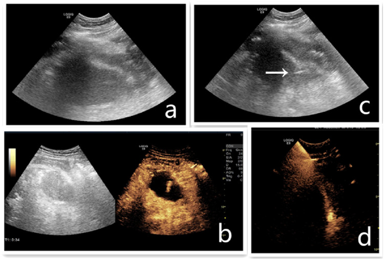
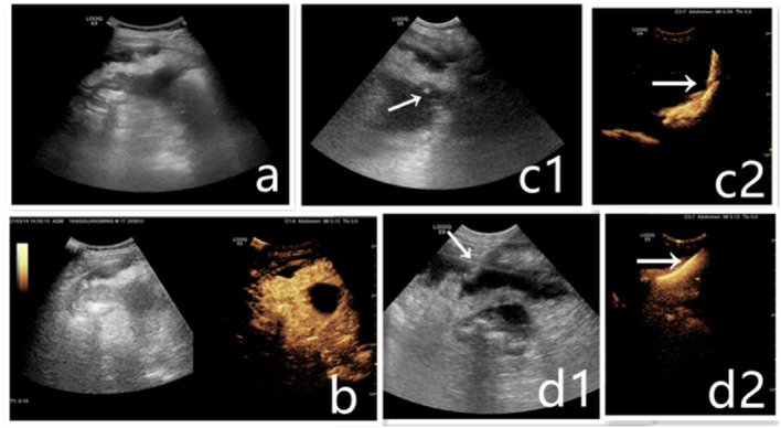
Results: An ultrasound contrast agent was used preoperatively to assess the extent of the abscess. Liquefaction and necrosis were observed in the abscess cavity. CEUS showed hyperenhancement in the wall of the abscess in the arterial phase and the liquefied necrotic area in the abscess cavity was not enhanced before PCD. CEUS allowed operators to confidently identify the puncture site. Amongst the 30 cases of PCD, 27 cases showed the clear positioning of the drainage tube. The head of the drainage tube was placed in the ideal position and development could be seen in the abscess cavity. The diffusion effect of the contrast agent was good with no spillover and the drainage was unobstructed. Abscess development was observed in 3 patients after puncture injection of the contrast agent but the head of the drainage tube was not in the predetermined position. After adjusting the position of the drainage tube, CEUS was repeated and showed a strong diffusion effect of the contrast agent.
Conclusions: Intravenous injection of Sonovue before PCD of periappendiceal abscesses can evaluate the extent of the abscess, liquefaction and necrosis in the abscess cavity. The approach can also provide guidance for the placement of the drainage tube. After the operation, a diluted contrast agent was injected through the drainage tube. The position of the drainage tube and the flow direction of the contrast agent could be seen. This approach has good value for clinical applications to accurately judge the position of the drainage tube.
Keywords: Contrast-enhanced ultrasound; Periappendiceal abscess (PCD).
© 2022. Società Italiana di Ultrasonologia in Medicina e Biologia (SIUMB).
Conflict of interest statement
None.
Figures
Similar articles
-
Intracavitary contrast-enhanced ultrasound in abscess drainage--feasibility and clinical value.Scand J Gastroenterol. 2016 Jan;51(1):41-7. doi: 10.3109/00365521.2015.1066423. Epub 2015 Jul 11. Scand J Gastroenterol. 2016. PMID: 26166454
-
Periappendiceal abscesses: percutaneous drainage.Radiology. 1987 Apr;163(1):23-6. doi: 10.1148/radiology.163.1.3823441. Radiology. 1987. PMID: 3823441
-
A Milestone: Approval of CEUS for Diagnostic Liver Imaging in Adults and Children in the USA.Ultraschall Med. 2016 Jun;37(3):229-32. doi: 10.1055/s-0042-107411. Epub 2016 Jun 8. Ultraschall Med. 2016. PMID: 27276056 English.
-
Interventional and surgical treatment of pancreatic abscess.World J Surg. 1997 Feb;21(2):162-8. doi: 10.1007/s002689900209. World J Surg. 1997. PMID: 8995072 Review.
-
Contrast-enhanced ultrasound imaging of the liver: a review of the clinical evidence for SonoVue and Sonazoid.Abdom Radiol (NY). 2020 Nov;45(11):3779-3788. doi: 10.1007/s00261-020-02573-9. Abdom Radiol (NY). 2020. PMID: 32424608 Review.
Cited by
-
Efficacy of Antibiotic Therapy Alone Versus Antibiotics With Percutaneous Drainage in Periappendiceal Abscess: A Systematic Review and Meta-Analysis.Cureus. 2024 Nov 19;16(11):e73979. doi: 10.7759/cureus.73979. eCollection 2024 Nov. Cureus. 2024. PMID: 39703257 Free PMC article. Review.
References
MeSH terms
Substances
LinkOut - more resources
Full Text Sources
Medical
