. 2022 Aug;298(8):102264.
doi: 10.1016/j.jbc.2022.102264. Epub 2022 Jul 14.

Phosphate position is key in mediating transmembrane ion channel TMEM16A-phosphatidylinositol 4,5-bisphosphate interaction

Affiliations

Phosphate position is key in mediating transmembrane ion channel TMEM16A-phosphatidylinositol 4,5-bisphosphate interaction

Maiwase Tembo et al. J Biol Chem. 2022 Aug.

Abstract

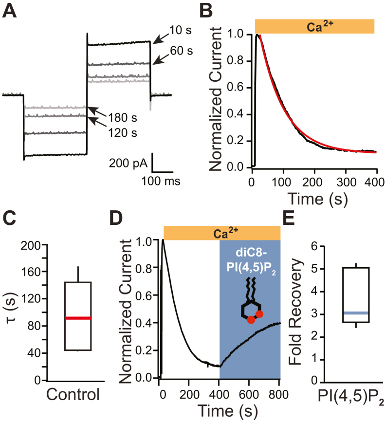
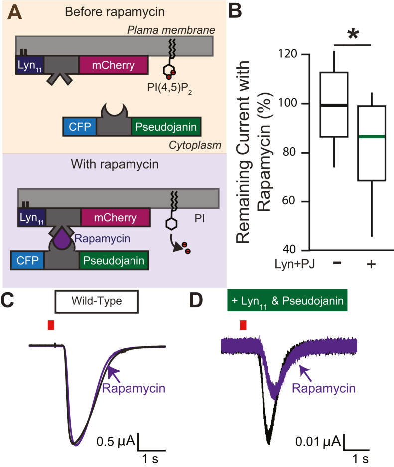
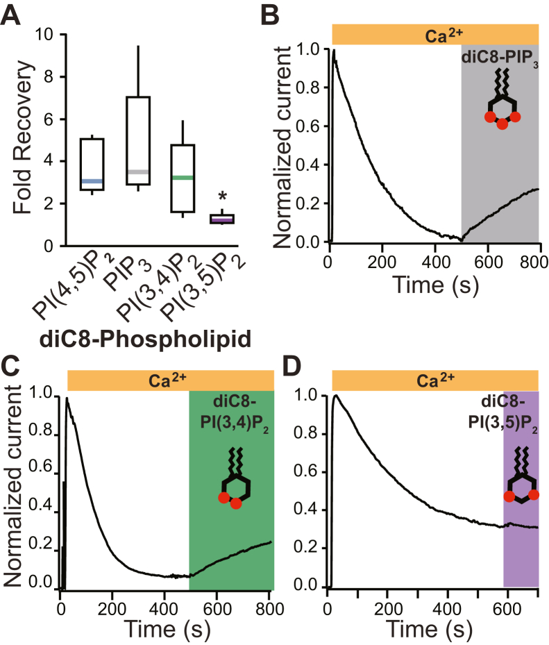
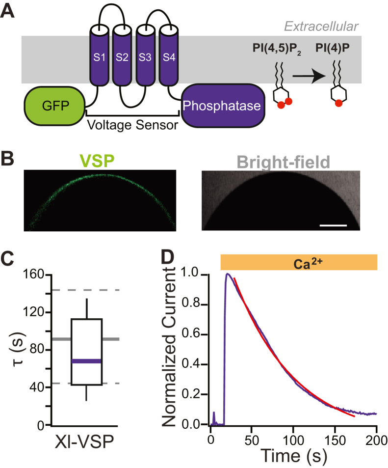
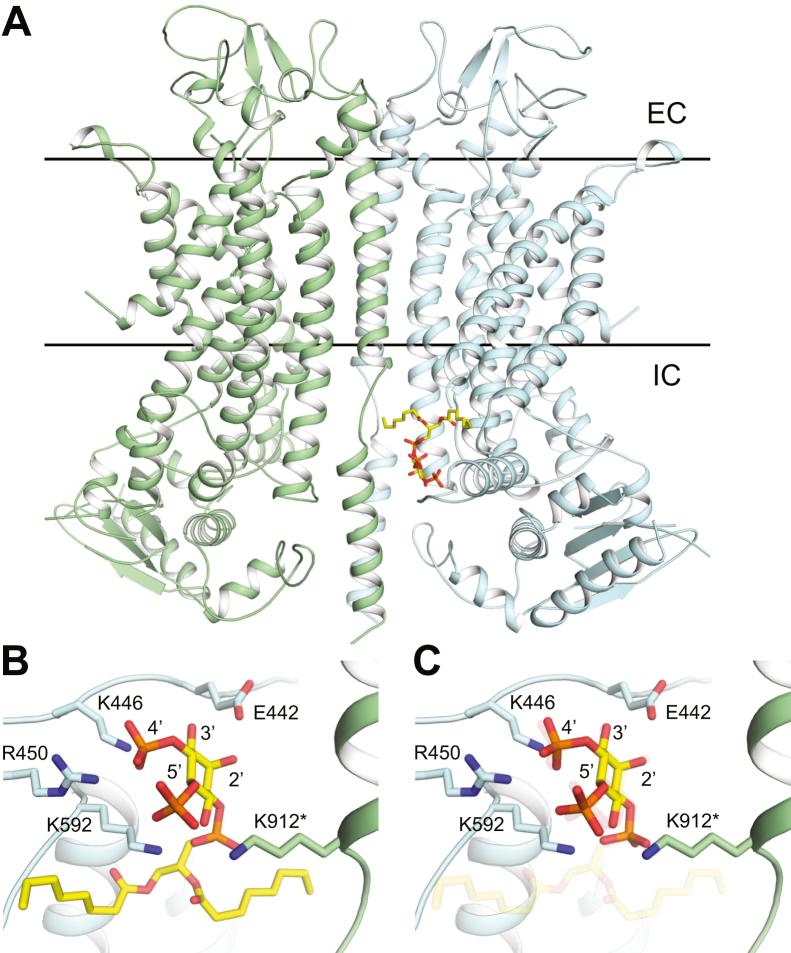
TransMEMbrane 16A (TMEM16A) is a Ca2+-activated Cl- channel that plays critical roles in regulating diverse physiologic processes, including vascular tone, sensory signal transduction, and mucosal secretion. In addition to Ca2+, TMEM16A activation requires the membrane lipid phosphatidylinositol 4,5-bisphosphate (PI(4,5)P2). However, the structural determinants mediating this interaction are not clear. Here, we interrogated the parts of the PI(4,5)P2 head group that mediate its interaction with TMEM16A by using patch- and two-electrode voltage-clamp recordings on oocytes from the African clawed frog Xenopus laevis, which endogenously express TMEM16A channels. During continuous application of Ca2+ to excised inside-out patches, we found that TMEM16A-conducted currents decayed shortly after patch excision. Following this rundown, we show that the application of a synthetic PI(4,5)P2 analog produced current recovery. Furthermore, inducible dephosphorylation of PI(4,5)P2 reduces TMEM16A-conducted currents. Application of PIP2 analogs with different phosphate orientations yielded distinct amounts of current recovery, and only lipids that include a phosphate at the 4' position effectively recovered TMEM16A currents. Taken together, these findings improve our understanding of how PI(4,5)P2 binds to and potentiates TMEM16A channels.

Keywords: anion channel; calcium; chloride channel; oocyte; patch clamp; phospholipid; signal transduction; transmembrane member 16A; xenopus.


Conflict of interest statement

Conflict of interest The authors declare that they have no conflicts of interest with the contents of this article.

Figures

Figure 1
Figure 1
TMEM16A Ca2+-evoked Clcurrents rundown in excised patches and are recovered by a diC8-PI(4,5)P2application.A, example currents recorded at the indicated times during 150 ms steps to −60 and +60 mV, recorded using excised inside–out macropatches from Xenopus laevis oocytes. B, normalized plot of current measured at −60 mV versus time, fit with a single exponential (red line). C, box plot distribution of the rate of current decay (τ), measured by fitting plots of relative current versus time with single exponentials (N = 11). The central line denotes the median, the box denotes 25 to 75% of the data, and the whiskers represent 10 to 90% of the data. D, a soluble synthetic analog of PI(4,5)P2, diC8-PI(4,5)P2, was applied to excised inside–out patches once current had stably rundown. Currents were recorded at −60 mV. E, box plot distribution of the fold current recovered after the application of diC8-PI(4,5)P2 with Ca2+ (N = 8). diC8-PI(4,5)P2, dioctanoyl phosphatidylinositol 4,5-bisphosphate; TMEM16A, TransMEMbrane 16A.
Figure 2
Figure 2
TMEM16A Ca2+-evoked Clcurrents are depleted in whole Xenopus laevis oocytes by dephosphorylation of PI(4,5)P2.A, schematic demonstrating pseudojanin translocation to the plasma membrane. To express pseudojanin at the membrane, the membrane tether Lyn11-mCherry and pseudojanin-CFP RNAs were both injected into X. laevis oocytes. Lyn11-mCherry expresses at the plasma membrane, and pseudojanin expresses in the cytoplasm. Upon rapamycin application, rapamycin binds Lyn11-mCherry and induces the membrane translocation of pseudojanin-CFP. Once at the membrane, pseudojanin-CFP dephosphorylates PI(4,5)P2 at the 4′ and 5′ position. The effects of pseudojanin on whole-cell TMEM16A Ca2+-evoked Cl currents were measured using the two-electrode voltage-clamp technique. B, box plot distribution of the percentage remaining current observed in uninjected control and pseudojanin-CFP–expressing X. laevis oocytes after incubation in 10 μM rapamycin for 5 min. The percent of remaining currents was significantly different (p = 0.02) as determined by a two-tailed t test. ∗ denotes p < 0.05. C and D, example of whole-cell currents recorded at −80 mV before and after rapamycin application in oocytes expressing pseudojanin-CFP. Current was recorded in control solution (black) and after incubation in rapamycin for 5 min (purple). Red bar represents 250 ms duration of UV light application. CFP, cyan fluorescent protein; PI(4,5)P2, phosphatidylinositol 4,5-bisphosphate; TMEM16A, TransMEMbrane 16A.
Figure 3
Figure 3
Phospholipid analogs differentially recovered TMEM16A. Soluble synthetic analogs of PIP3 (diC8-PIP3), PI(3,4)P2 (diC8-PI(3,4)P2), and PI(3,5)P2 were applied to excised inside–out patches once current had stably rundown. Currents were recorded at −60 mV. A, box plot distribution of the fold current recovered after the application of diC8-PI(4,5)P2 (N = 9), diC8-PIP3 (N = 7), diC8-PI(3,4)P2 (N = 6), or diC8-PI(3,5)P2 (N = 5). Representative plots of normalized currents versus time, before and during application of 100 μM diC8-PIP3 (B), diC8-PI(3,4)P2 (C), or diC8-PI(3,5)P2 (D). ∗ represents p < 0.025 as determined by ANOVA and Tukey's HSD post hoc tests. diC8-PI(3,4)P2, dioctanoyl phosphatidylinositol 3,4-bisphosphate; diC8-PI(3,5)P2, dioctanoyl phosphatidylinositol 3,5-bisphosphate; diC8-PI(4,5)P2, dioctanoyl phosphatidylinositol 4,5-bisphosphate; diC8-HSD, honestly significant difference; PIP3, dioctanoyl phosphatidylinositol 3,4,5-trisphosphate; PI(3,4)P2, phosphatidylinositol 3,4-bisphosphate; PI(3,5)P2, dioctanoyl phosphatidylinositol 3,5-bisphosphate; PIP3, phosphatidyl 3,4,5-trisphosphate; TMEM16A, TransMEMbrane 16A.
Figure 4
Figure 4
Phospholipids with phosphates at position 4′ of the inositol ring recover current. Soluble synthetic analogs of PI3P, PI4P, and PI5P were applied to excised inside–out patches once current had stably rundown. Currents were recorded at −60 mV. Representative plots of normalized currents versus time, before and during application of 100 μM diC8-PI3P (A), 100 μM diC8-PI5P (B), or 100 μM diC8-PI4P (D). C, box plot distribution of the fold current recovered after the application of diC8-PI(4,5)P2 (N = 9), diC8-PI3P (N = 7), diC8-PI4P (N = 7), or diC8-PI5P (N = 5). ∗ denotes p < 0.05 between indicated treatment and diC8-PI(4,5)P2, as determined by ANOVA and Tukey’s HSD post hoc tests. diC8-PI3P, dioctanoyl 3-monophosphate; diC8-PI4P, dioctanoyl 4-monophosphate; diC8-PI5P, dioctanoyl 5-monophosphate; HSD, honestly significant difference.
Figure 5
Figure 5
Xl-VSP does not significantly change TMEM16A current rundown. Inside–out patch-clamp recordings were conducted on macropatches excised from Xenopus laevis oocytes expressing Xl-VSP. A, schematic depicting a VSP tagged with GFP. B, confocal and bright-field images of a representative X. laevis oocyte expressing GFP-tagged Xl-VSP at the plasma membrane. Bar denotes 200 μm. C, box plot distribution of the rate of current decay (τ), measured by fitting plots of relative current versus time with single exponentials for the Xl-VSP expressing (purple) (N = 6). Background gray dashed lines denote 25 to 75% of the data spread, and the solid line represents the median rate of rundown measured from patches recorded under the control conditions (plotted in Fig. 1C). D, representative plot of normalized currents versus time following VSP activation. TMEM16A, TransMEMbrane 16A; Xl-VSP, Xenopus laevis voltage-sensing phosphatase.
Figure 6
Figure 6
Docking suggests key PI(4,5)P2phosphate interactions with TMEM16A. Docking was performed with either diC8-PI(4,5)P2 or IP3 into a homology model of Xenopus laevis TMEM16A (xTMEM16A). A, position of diC8-PI(4,5)P2 shown against the homology model of xTMEM16A. Lines indicating the position of the intracellular and extracellular boundaries of the plasma membrane were created using the OPM entry for mouse TMEM16A (PDB: 5OYB). B, detailed view of the hypothesized PI(4,5)P2–xTMEM16A interaction. Interacting residues (E442, K446, R450, K592, and K912 from the other chain) and phosphates (positions 2′–5′) are highlighted. C, superposition of docked IP3 (foreground) on the PI(4,5)P2–xTMEM16A (transparency) interaction. diC8-PI(4,5)P2, dioctanoyl phosphatidylinositol 4,5-bisphosphate; PDB, Protein Data Bank; PI(4,5)P2, dioctanoyl phosphatidylinositol 4,5-bisphosphate; TMEM16A, TransMEMbrane 16A; xTMEM16A, Xenopus laevis TransMEMbrane 16A.

References

    1. Askew Page H.R., Dalsgaard T., Baldwin S.N., Jepps T.A., Povstyan O., Olesen S.P., et al. TMEM16A is implicated in the regulation of coronary flow and is altered in hypertension. Br. J. Pharmacol. 2019;176:1635–1648. - PMC - PubMed
    1. Huang F., Wong X., Jan L.Y. International union of basic and clinical pharmacology. LXXXV: calcium-activated chloride channels. Pharmacol. Rev. 2012;64:1–15. - PMC - PubMed
    1. Pedemonte N., Galietta L.J. Structure and function of TMEM16 proteins (anoctamins) Physiol. Rev. 2014;94:419–459. - PubMed
    1. Jin X., Shah S., Liu Y., Zhang H., Lees M., Fu Z., et al. Activation of the Cl- channel ANO1 by localized calcium signals in nociceptive sensory neurons requires coupling with the IP3 receptor. Sci. Signal. 2013;6:ra73. - PMC - PubMed
    1. Liu B., Linley J.E., Du X., Zhang X., Ooi L., Zhang H., et al. The acute nociceptive signals induced by bradykinin in rat sensory neurons are mediated by inhibition of M-type K+ channels and activation of Ca2+-activated Cl- channels. J. Clin. Invest. 2010;120:1240–1252. - PMC - PubMed

Publication types

MeSH terms