Review
. 2023 Feb:132:39-55.
doi: 10.1016/j.humpath.2022.07.006. Epub 2022 Jul 16.

COVID-19: gastrointestinal and hepatobiliary manifestations

Affiliations
Review

COVID-19: gastrointestinal and hepatobiliary manifestations

Angela R Shih et al. Hum Pathol. 2023 Feb.

Abstract

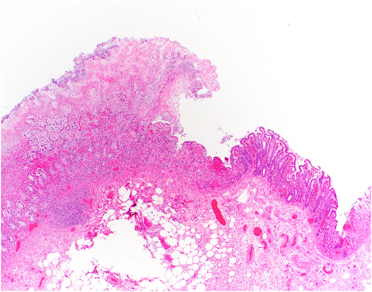
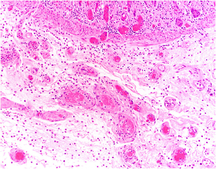
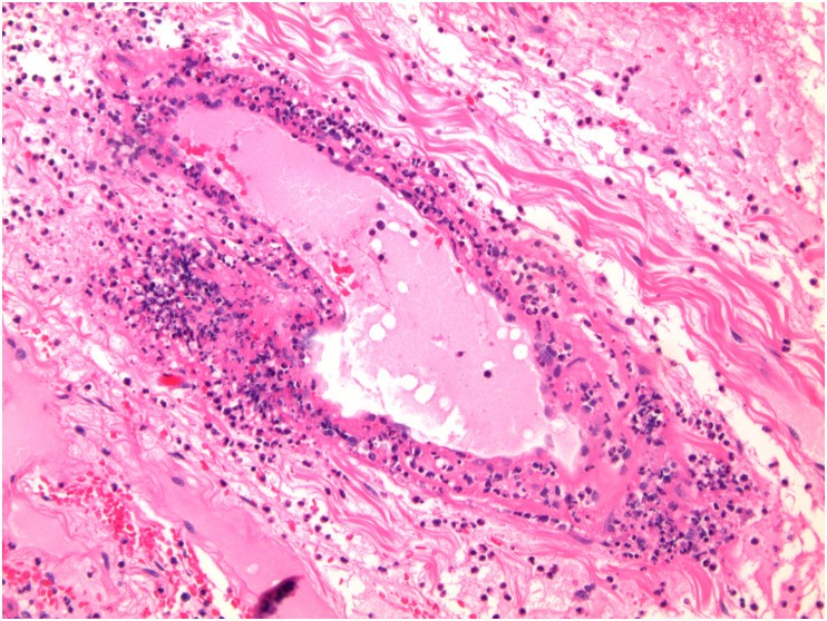
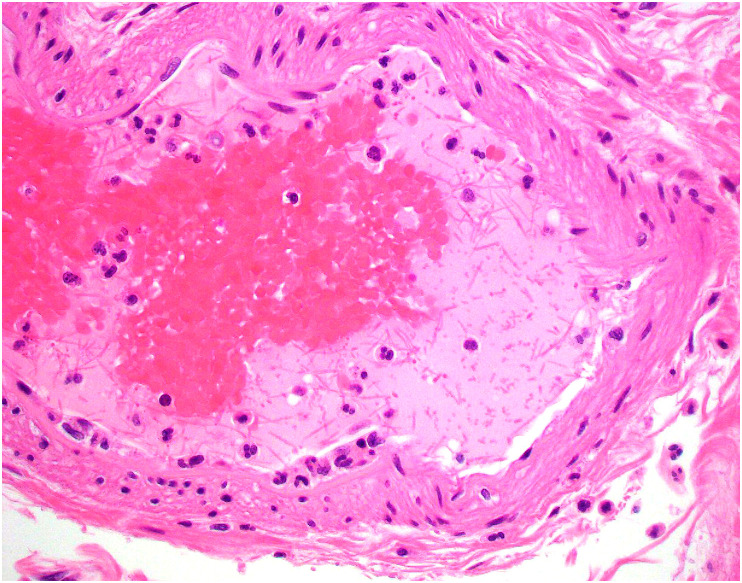
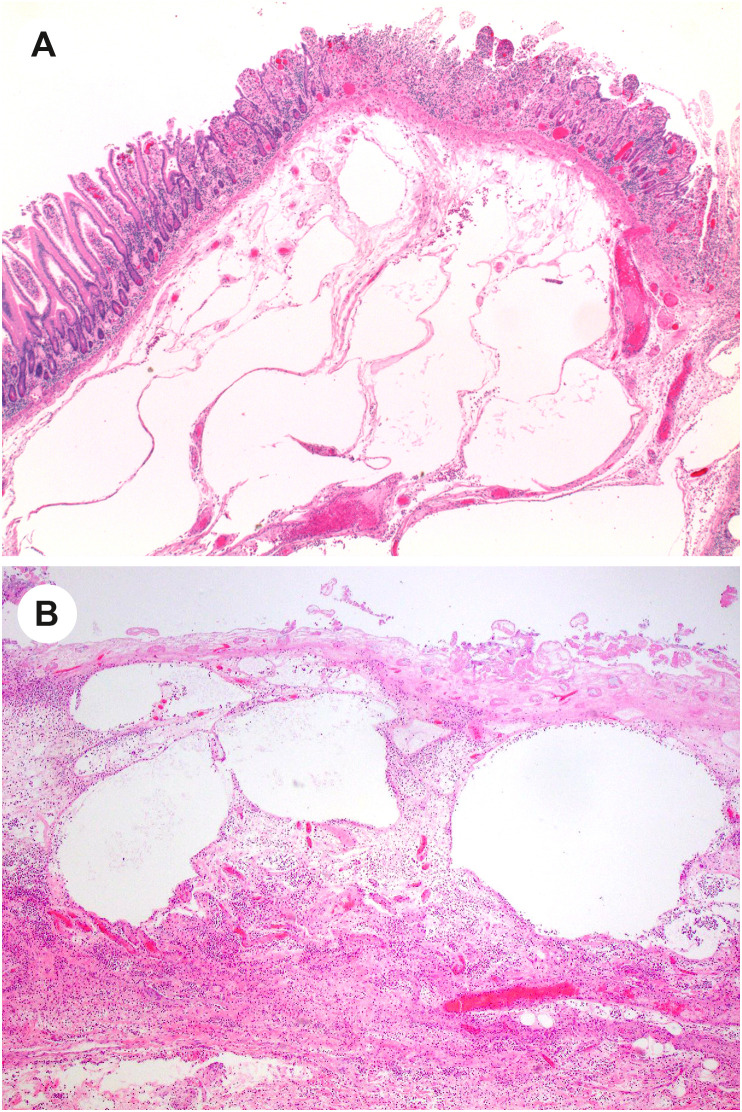
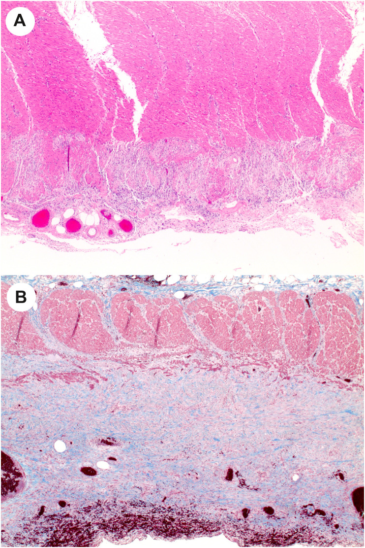
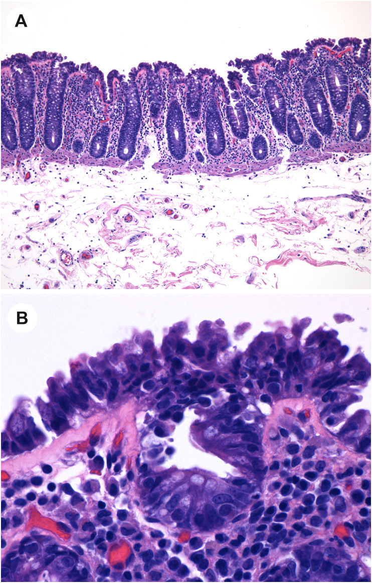
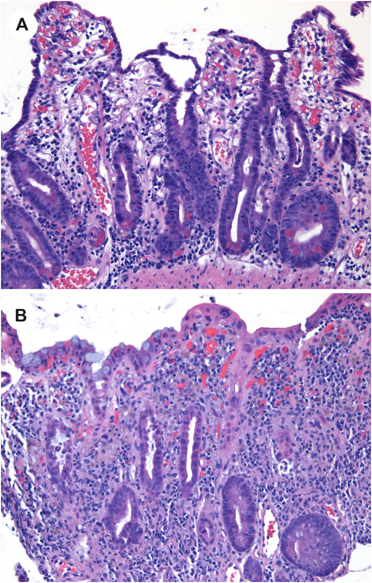
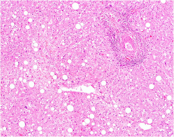
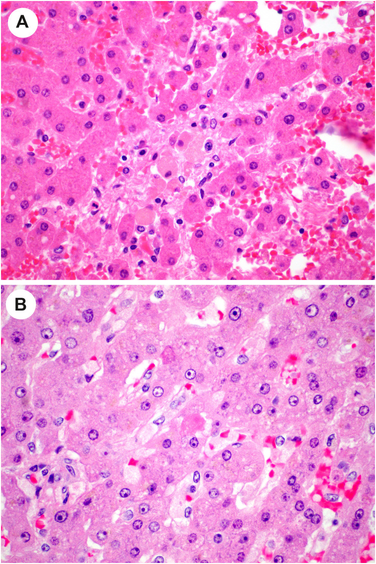
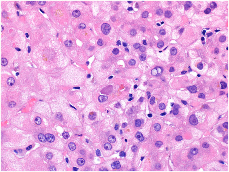
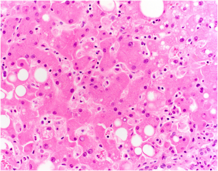
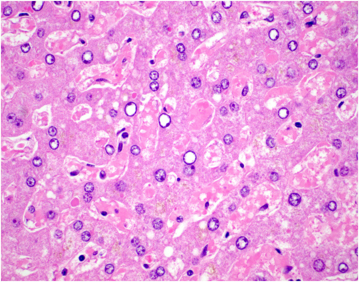
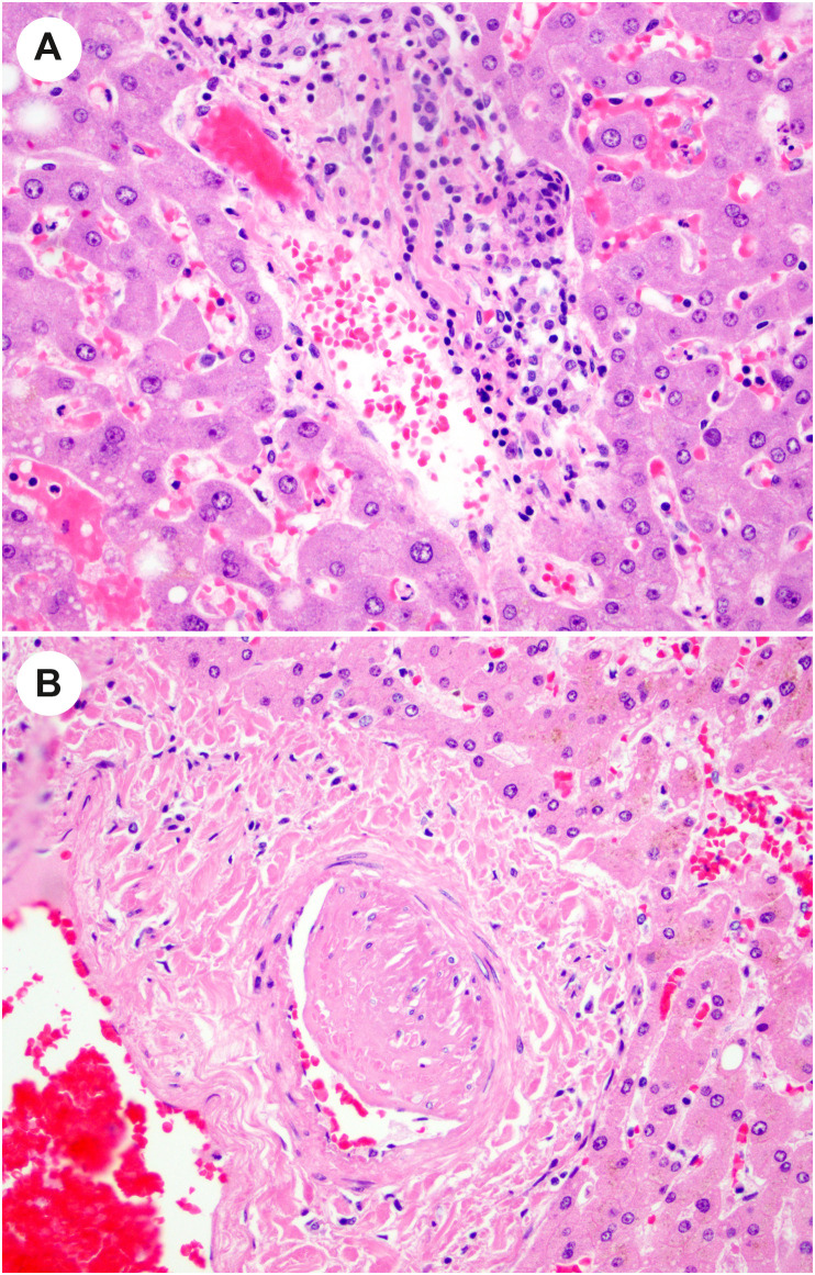
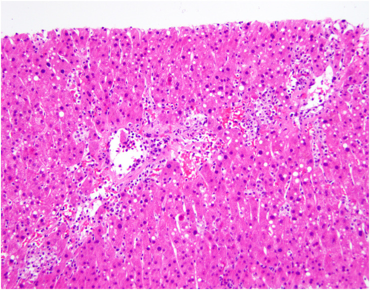
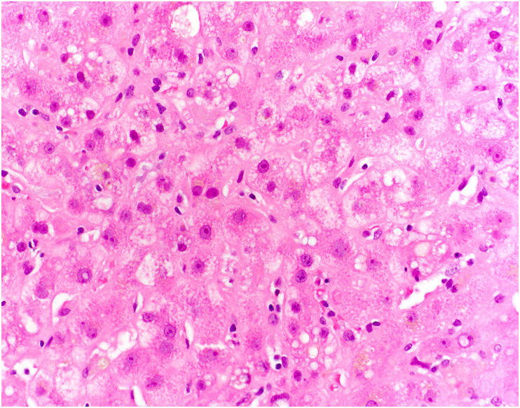
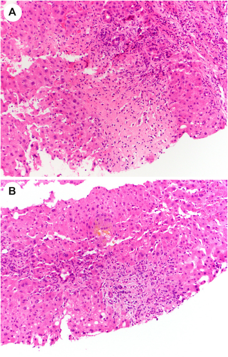
SARS-CoV-2 is the viral agent of COVID-19, a pandemic that surfaced in 2019. Although predominantly a respiratory ailment, patients with COVID-19 can have gastrointestinal (GI) and hepatobiliary manifestations. These manifestations are often mild and transient, but they can be severe and consequential. In the GI tract, ischemic enterocolitis is the most common and significant consequence of COVID-19. In the liver, the reported pathologic findings may often be related to consequences of severe systemic viral infection, but reports of hepatitis presumed to be due to SARS-CoV-2 suggest that direct viral infection of the liver may be a rare complication of COVID-19. In both the GI tract and liver, lingering symptoms of GI or hepatic injury after resolution of pulmonary infection may be part of the evolving spectrum of long COVID.

Keywords: COVID-19; Colon; Gastrointestinal; Liver; Pathology.


Figures

Fig. 1
Fig. 1
Ischemic enterocolitis in COVID-19. Low-power view of a colon specimen shows mucosal necrosis with a fibrinopurulent exudate (left) with a sharp transition to viable mucosa (right). The specimens also show congestion, submucosal edema, and epithelial injury characteristic of ischemic enterocolitis.
Fig. 2
Fig. 2
Fibrin thrombi in COVID-19–related ischemic enterocolitis. Small vessels within the submucosa beneath necrotic mucosa shows fibrin thrombi in small vessels.
Fig. 3
Fig. 3
Perivascular neutrophils in the submucosa in COVID-19–related ischemic enterocolitis. Vessels within the submucosa may show changes reminiscent of vasculitis, with perivascular neutrophils and, in some cases, degeneration of the wall. (Courtesy of Dr. M. Lisa Zhang).
Fig. 4
Fig. 4
Fibrin strands in large submucosal vessels in COVID-19–related ischemic enterocolitis. In some cases of COVID-19–related ischemic bowel, large submucosal vessels show fibrin strands within stagnant blood. This may be a reflection of hypercoagulability.
Fig. 5
Fig. 5
Pneumatosis intestinalis in COVID-19–related ischemic enterocolitis. A and B, Two cases that demonstrate large “empty” spaces in the submucosa in ischemic enterocolitis that are compatible with pneumatosis intestinalis. (Courtesy of Dr. M. Lisa Zhang).
Fig. 6
Fig. 6
Muscularis propria attenuation in COVID-19–related ischemic enterocolitis. A, Rare cases of COVD-19 ischemic bowel show changes that are typically seen in chronic ischemic, such as attenuation of the external layer of the muscularis propria. B, Trichrome stain of a different case shows fibrous replacement of the external layer of the muscularis propria, as well as subserosal fibrosis. (Courtesy of Dr. M. Lisa Zhang).
Fig. 7
Fig. 7
Mucosal injury in COVID-19. A, Low-power view of colonic mucosa shows tufting of the superficial epithelium. B, High-power view shows that the enterocytes are injured with disarray, hyperchromatic disordered nuclei, epithelial tufting, and amphophilic cytoplasm. (Courtesy of Dr. Rhonda Yantiss).
Fig. 8
Fig. 8
Widespread mucosal injury in COVID-19. A, Small intestinal biopsy in a patient with COVID-19 demonstrates villous blunting with cuboidalization and attenuation of the duodenal enterocytes, and crypt hyperplasia. B, A biopsy of the colon in the same patient shows cuboidal, disarrayed colonocytes with attenuated crypts and increased cellularity of the lamina propria. A few crypts (such as the one at left) show sloughed epithelial cells within the crypt. (Courtesy of Dr. Rhonda Yantiss).
Fig. 9
Fig. 9
Steatosis in COVID-19. An autopsy from a patient who died of COVID-19 demonstrates patchy steatosis, including large droplet fat and scattered smaller droplets within hepatocytes, without strict zonal predilection.
Fig. 10
Fig. 10
Lobular injury in autopsy livers from patients with COVID-19. A, Focal lobular necrotic focus with pigmented macrophages and hepatocyte dropout. B, In this view, an acidophil body is noted (center) as well as increased histiocytes in sinusoids. (Courtesy of Dr. Stephen Lagana).
Fig. 11
Fig. 11
Cholestasis in autopsy liver from patient with COVID-19. High-power view shows enlarged reactive-appearing hepatocytes with feathery degeneration, and cytoplasmic pigment consistent with intracellular cholestasis. (Courtesy of Dr. Michael Torbenson).
Fig. 12
Fig. 12
Kupffer cell hyperplasia in autopsy liver from patient with COVID-19. In this high-power view, the hepatic plates are somewhat disarrayed, and the sinusoids are expanded with numerous histiocytes and scattered inflammatory cells. Mild steatosis is also present.
Fig. 13
Fig. 13
Thrombotic changes in the liver in patients who died of COVID-19. The lobule in this liver shows numerous fibrin thrombi within the sinusoids. (Courtesy of Dr. Stephen Lagana).
Fig. 14
Fig. 14
Vascular changes in autopsy livers in patients with COVID-19. A, In this portal tract, there is mild inflammation of the portal vein. B, A hypertrophied artery is present in this view of a portal tract. (Courtesy of Dr. Stephen Lagana).
Fig. 15
Fig. 15
Hepatitis in a liver allograft in a patient with COVID-19. The lobule shows mononuclear inflammation with hepatocyte dropout, a few acidophil bodies, and small droplet steatosis in zone 3. Elsewhere, the portal changes suggested acute cellular rejection. (Courtesy of Dr. Stephen Lagana).
Fig. 16
Fig. 16
Post–COVID-19 cholangiopathy. In the lobule in this liver biopsy, the hepatocytes are enlarged, reactive appearing, and demonstrate feathery degeneration. There is also cytoplasmic pigment, consistent with intrahepatic cholestasis.
Fig. 17
Fig. 17
Portal obstructive type changes in post–COVID-19 cholangiopathy. A, A portal tract (upper center) shows inflammation and ductular reaction. Below the portal tract (center), there is an area of hepatocyte dropout suggestive of a bile infarct. B, In this portal tract, there is ductular reaction and periportal lobular cholestasis.

References

    1. Zhang H., Liao Y.-S., Gong J., et al. Clinical characteristics of coronavirus disease (COVID-19) patients with gastrointestinal symptoms: a report of 164 cases. Dig Liver Dis. 2020;52:1076–1079. doi: 10.1016/j.dld.2020.04.034. - DOI - PMC - PubMed
    1. Hajifathalian K., Krisko T., Mehta A., et al. Gastrointestinal and hepatic manifestations of 2019 novel coronavirus disease in a large cohort of infected patients from New York: clinical implications. Gastroenterology. 2020;159:1137–1140. doi: 10.1053/j.gastro.2020.05.010. e2. - DOI - PMC - PubMed
    1. Elmunzer B.J., Spitzer R.L., Foster L.D., et al. Digestive manifestations in patients hospitalized with coronavirus disease 2019. Clin Gastroenterol Hepatol. 2021;19:1355–1365. doi: 10.1016/j.cgh.2020.09.041. e4. - DOI - PMC - PubMed
    1. Cao T.-T., Zhang G.-Q., Pellegrini E., et al. COVID-19 and its effects on the digestive system. World J Gastroenterol. 2021;27:3502–3515. doi: 10.3748/wjg.v27.i24.3502. - DOI - PMC - PubMed
    1. Cheung K.S., Hung I.F.N., Chan P.P.Y., et al. Gastrointestinal manifestations of SARS-CoV-2 infection and virus load in fecal samples from a Hong Kong cohort: systematic review and meta-analysis. Gastroenterology. 2020;159:81–95. doi: 10.1053/j.gastro.2020.03.065. - DOI - PMC - PubMed