Review
. 2022 Sep:80:101687.
doi: 10.1016/j.arr.2022.101687. Epub 2022 Jul 16.

SARS-CoV-2 and the central nervous system: Emerging insights into hemorrhage-associated neurological consequences and therapeutic considerations

Affiliations
Review

SARS-CoV-2 and the central nervous system: Emerging insights into hemorrhage-associated neurological consequences and therapeutic considerations

Joy Mitra et al. Ageing Res Rev. 2022 Sep.

Abstract

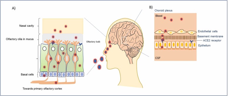
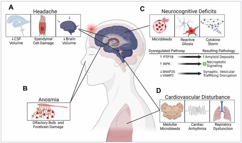
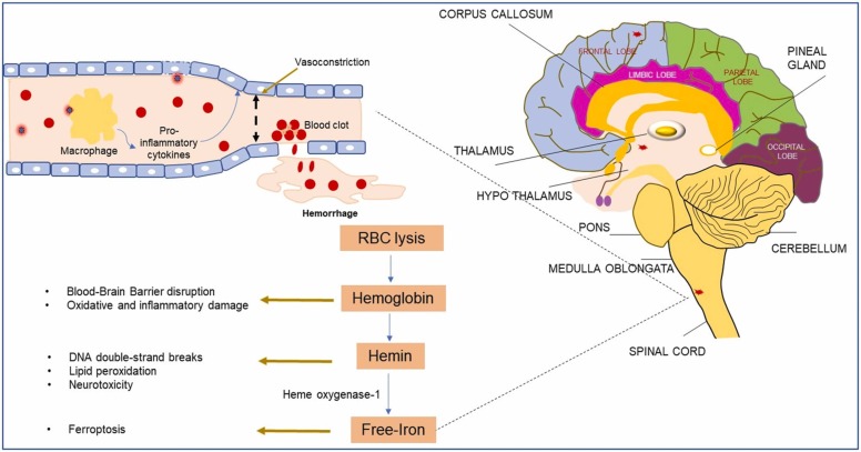
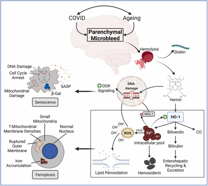
Coronavirus disease 2019 (COVID-19), caused by the Severe Acute Respiratory Syndrome Coronavirus 2 (SARS-CoV-2) continues to impact our lives by causing widespread illness and death and poses a threat due to the possibility of emerging strains. SARS-CoV-2 targets angiotensin-converting enzyme 2 (ACE2) before entering vital organs of the body, including the brain. Studies have shown systemic inflammation, cellular senescence, and viral toxicity-mediated multi-organ failure occur during infectious periods. However, prognostic investigations suggest that both acute and long-term neurological complications, including predisposition to irreversible neurodegenerative diseases, can be a serious concern for COVID-19 survivors, especially the elderly population. As emerging studies reveal sites of SARS-CoV-2 infection in different parts of the brain, potential causes of chronic lesions including cerebral and deep-brain microbleeds and the likelihood of developing stroke-like pathologies increases, with critical long-term consequences, particularly for individuals with neuropathological and/or age-associated comorbid conditions. Our recent studies linking the blood degradation products to genome instability, leading to cellular senescence and ferroptosis, raise the possibility of similar neurovascular events as a result of SARS-CoV-2 infection. In this review, we discuss the neuropathological consequences of SARS-CoV-2 infection in COVID survivors, focusing on possible hemorrhagic damage in brain cells, its association to aging, and the future directions in developing mechanism-guided therapeutic strategies.

Keywords: Brain fog; COVID-19; Coronavirus; Ferroptosis; Genome instability; Hemorrhage; SARS-CoV-2; Senescence.


Conflict of interest statement

The authors declare that the study was conducted in the absence of any commercial or financial relationships that could be construed as a potential conflict of interest.

Figures

Fig. 1
Fig. 1
The interaction of SARS-CoV-2 with olfactory cilia for entering the central nervous system (CNS). A) Different layers of cells in the nasal cavity that contain receptors for the very first viral entry into the human host. Some of the olfactory cells express ACE2 receptors that endocytose bound viral particles, before their way to the olfactory bulb in the brain. B) Schematic of the cellular barrier between cerebrospinal fluid and circulating blood in the choroid plexus region. Both endothelial and basement epithelial cells express the ACE2 receptors facilitating the internalization of virions in the brain fluid system.
Fig. 2
Fig. 2
SARS-CoV-2-associated long-COVID pathogenic mechanisms in the COVID-19 survivors. A) Chronic headaches can be triggered due to the damage of ependymal cells, reduced cerebrospinal fluid (CSF) content, and the resulting shrunken brain volume. B) Viral infection-induced neuronal damage in the olfactory bulb and forebrain areas leads to acute anosmia. C) Initiation of neurocognitive decline resulting from the hemorrhagic microbleed-induced reactive gliosis, neuroimmune activation, and cytokine storms. Several signaling cascades related to synaptic transmission, vesicular trafficking, amyloidogenesis, and necroptotic pathways are dysregulated in this process. D) Similar mechanisms associated with the microbleeds in the deeper brain regions like medullar oblongata lead to cardiac arrhythmia and abnormal respiratory functions.
Fig. 3
Fig. 3
SARS-CoV-2 infection-induced microbleeds in the brain and its potential consequences. A) Mechanism of virion recognition by endothelial ACE2 receptors, which in turn induce activation of a cytokine storm, leading to vessel rupture and downstream genotoxic and cytotoxic effects. B) Illustration of different brain regions in the sagittal section, indicating the formation of microbleeds as red spots in certain locations.
Fig. 4
Fig. 4
Pathomechanistic implications of ferroptosis in age-related neuronal damage in the long COVID-19 survivors. Microbleeds are frequently detected in the aged brain in neuroimaging examinations. However, COVID-19 drastically increases the number of these microhemorrhagic lesions in the brain parenchyma. Following the hemolysis, hemoglobin disintegrates into globin and hemin parts. The schematic of ferroptosis illustrates how hemin sequentially undergoes biodegradation to form the pro-oxidant free iron ions, which in turn get engaged in Fenton’s reaction to produce reactive oxygen species (ROS), resulting in the oxidative stress-induced genome damage as well as membrane disruption via lipid peroxidation mechanism, and ultimately cell death via ferroptosis. Since acutely overloaded intracellular iron (III) ions can inactivate oxidative DNA damage repair proteins like NEIL1, unrepaired DNA damage persistently sends out damage response signals, leading to cellular senescence. The key cellular hallmarks of a senescent and ferroptotic cell are indicated in the respective illustrations.

Similar articles

Cited by

References

    1. Ahn J.H., Kim J., Hong S.P., Choi S.Y., Yang M.J., Ju Y.S., Kim Y.T., Kim H.M., Rahman M.D.T., Chung M.K., Hong S.D., Bae H., Lee C.S., Koh G.Y. Nasal ciliated cells are primary targets for SARS-CoV-2 replication in the early stage of COVID-19. J. Clin. Investig. 2021;131(13) doi: 10.1172/jci148517. - DOI - PMC - PubMed
    1. Ahn M., Anderson D.E., Zhang Q., Tan C.W., Lim B.L., Luko K., Wen M., Chia W.N., Mani S., Wang L.C., Ng J.H.J., Sobota R.M., Dutertre C.A., Ginhoux F., Shi Z.L., Irving A.T., Wang L.F. Dampened NLRP3-mediated inflammation in bats and implications for a special viral reservoir host. Nat. Microbiol. 2019;4(5):789–799. doi: 10.1038/s41564-019-0371-3. - DOI - PMC - PubMed
    1. Akoudad S., Wolters F.J., Viswanathan A., de Bruijn R.F., van der Lugt A., Hofman A., Koudstaal P.J., Ikram M.A., Vernooij M.W. Association of cerebral microbleeds with cognitive decline and dementia. JAMA Neurol. 2016;73(8):934–943. doi: 10.1001/jamaneurol.2016.1017. - DOI - PMC - PubMed
    1. Albuquerque S., Teixeira A.M., Rocha J.C. COVID-19 and disenfranchised grief. Front. Psychiatry. 2021;12(114) doi: 10.3389/fpsyt.2021.638874. - DOI - PMC - PubMed
    1. Ali K.M., Ali A.M., Tawfeeq H.M., Figueredo G.P., Rostam H.M. Hypoalbuminemia in patients following their recovery from severe coronavirus disease 2019. J. Med. Virol. 2021;93(7):4532–4536. doi: 10.1002/jmv.27002. - DOI - PMC - PubMed

Publication types