

Evaluation of factors related to morphological masseter muscle changes after preoperative orthodontic treatment in female patients with skeletal class III dentofacial deformities
- PMID: 35843934
- PMCID: PMC9288706
- DOI: 10.1186/s12903-022-02319-7
Evaluation of factors related to morphological masseter muscle changes after preoperative orthodontic treatment in female patients with skeletal class III dentofacial deformities
Abstract
Background: The purpose of the current study was to investigate factors related to morphological changes in the masseter muscle after preoperative orthodontic treatment in patients with skeletal class III dentofacial deformities for analysis of muscle changes and malocclusions.
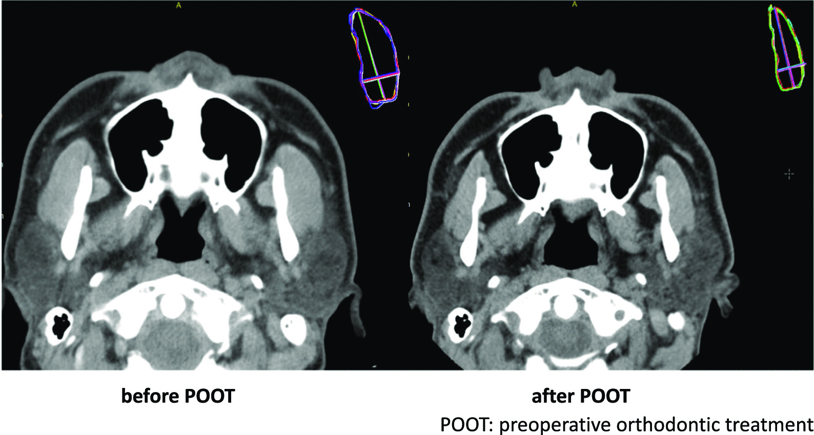
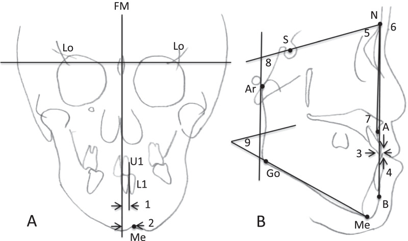
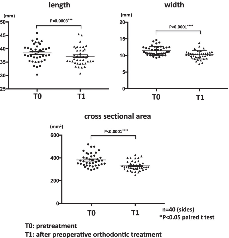
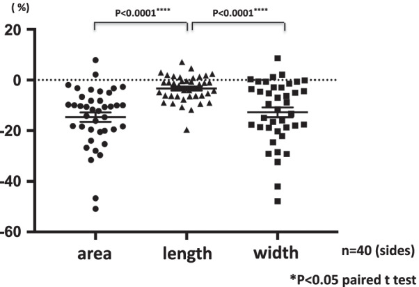
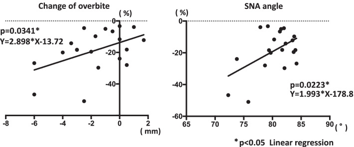
Methods: Twenty female patients with dentofacial deformities were included in the study. Computed tomography was performed before and after preoperative orthodontic treatment, and the lengths, widths, and cross-sectional areas of the masseter muscles were measured. Changes in these parameters were evaluated, and factors related to changes in masseter muscle area after preoperative orthodontic treatment were analyzed.
Results: The lengths, widths, and areas of masseter muscles were significantly smaller after preoperative orthodontic treatment. Smaller masseter muscle area was significantly associated with changes in overbite and pretreatment values of SNA angle.
Conclusions: Atrophy of the masseter muscle during preoperative orthodontic treatment was greater in patients with increased open bite due to improved dental compensation in patients with skeletal class III dentofacial deformities with maxillary retraction.
Keywords: Morphological changes of masseter muscle; Preoperative surgical orthodontic treatment; Skeletal class III dentofacial deformities.
© 2022. The Author(s).
Conflict of interest statement
The authors declare that they have no competing interests.
Figures
References
-
- Ueki K, Okabe K, Mukozawa A, et al. Assessment of ramus, condyle, masseter muscle, and occlusal force before and after sagittal split ramus osteotomy in patients with mandibular prognathism. Oral Surg Oral Med Oral Pathol Oral Radiol Endodontol. 2009;108(5):679–686. doi: 10.1016/j.tripleo.2009.05.042. - DOI - PubMed
MeSH terms
LinkOut - more resources
Full Text Sources
Research Materials
