. 2022 Jul 17;22(1):230.
doi: 10.1186/s12935-022-02639-2.

A pan-cancer analysis of thioredoxin-interacting protein as an immunological and prognostic biomarker

Affiliations

A pan-cancer analysis of thioredoxin-interacting protein as an immunological and prognostic biomarker

Xuxue Guo et al. Cancer Cell Int. .

Abstract

Background: The critical role of thioredoxin-interacting protein (TXNIP) in cellular sulfhydryl redox homeostasis and inflammasome activation is already widely known, however, no pan-cancer analysis is currently available.

Methods: We thus first explored the potential roles of TXNIP across thirty-three tumors mainly based on The Cancer Genome Atlas and Gene Expression Omnibus datasets.

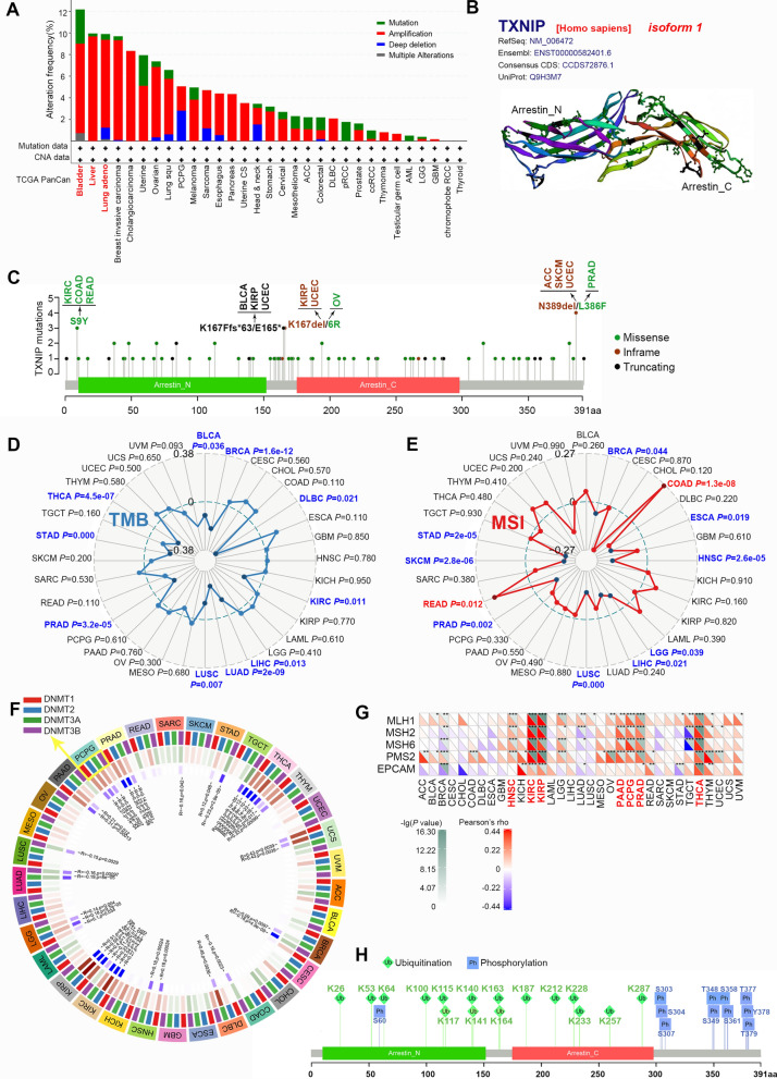
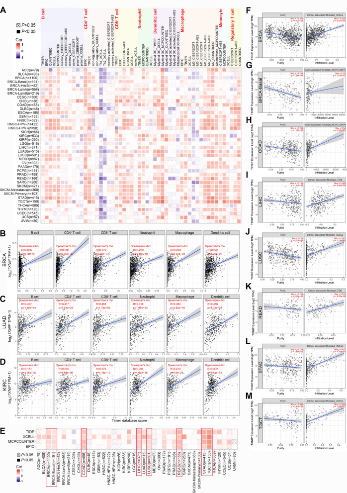
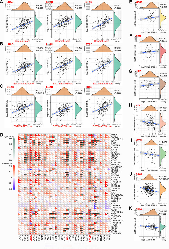
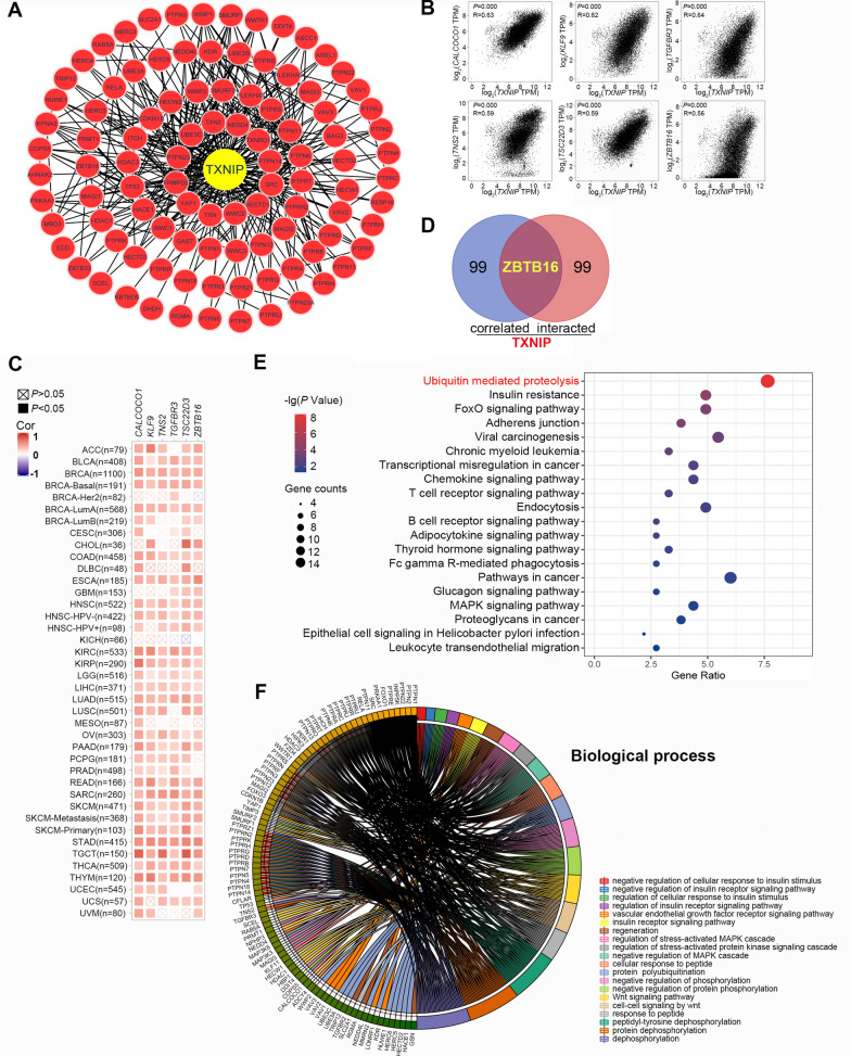
Results: TXNIP is lowly expressed in most cancers, and distinct associations exist between TXNIP expression and the prognosis of tumor patients. TXNIP expression was associated with tumor mutational burden, microsatellite instability, mismatch repair genes, tumor infiltrating immune cell abundance as well as cancer-associated fibroblasts. Moreover, ubiquitin mediated proteolysis, protein post-translational modification and other related pathways were involved in the functional mechanisms of TXNIP.

Conclusions: Our first pan-cancer study comprehensively revealed the carcinostatic role of TXNIP across different tumors. And this molecule may be considered as a potential immunological and prognostic biomarker.

Keywords: Genetic alteration; Immune infiltration; Pan-cancer; Prognosis; TXNIP; Ubiquitination.


Conflict of interest statement

The authors declare that the research was conducted in the absence of any commercial or financial relationships that could be construed as a potential conflict of interest.

Figures

Fig. 1
Fig. 1
TXNIP expression in different types of human tumors. A Diagram for the reported association between TXNIP and various tumors. The reported pathways activated by TXNIP in different tumors are presented in a pictorial manner. The relevant references are also indicated. B Increased or decreased TXNIP in data sets of different tumors compared to normal tissues in the Oncomine database. The cell number represents the dataset number that meets all thresholds, red represents over-expression, and blue represents under-expression. C Expression level of TXNIP in different types of human tumors from TCGA data via TIMER2. D For ACC, DLBC, BRCA, OV, SKCM and UCS in the TCGA project, the corresponding normal tissues in GTEx database were setted as controls. E TXNIP total protein in normal tissue and primary tissue of breast cancer, colon cancer, ovarian cancer, LUAD and UCEC in CPTAC dataset. (*P < 0.05; **P < 0.01; ***P < 0.001). RT-qPCR analysis of the TXNIP mRNA expression in tumor specimens and matched normal tissues from patients with F CRC (n = 50), G liver cancer (n = 32) and H BRCA (n = 10). I Western blotting analysis of the TXNIP expression in 18 pairs of randomly selected CRC samples. J Western blotting analysis of the TXNIP expression in 5 pairs of BRCA and 4 pairs of liver cancer samples. K Representative IHC staining of TXNIP in cancer and adjacent normal tissue from CRC patients. Scale bars: 100 μm. L Immunohistochemical score of TXNIP
Fig. 2
Fig. 2
Survival curves comparing the high and low expression of TXNIP in different tumors. A OS analyses in GEPIA2. B DFS analyses in GEPIA2. CF OS, RFS, PPS and DMFS survival curves of breast cancer in Kaplan–Meier plotter databases (n = 1402, n = 3955, n = 414, and n = 1805, respectively). GJ OS, RFS, PFS and DSS survival curves of liver cancer in Kaplan Meier plotter databases (n = 364, n = 316, n = 370, and n = 362, respectively). KM OS, PPS and FP survival curves of lung cancer in Kaplan Meier plotter databases (n = 1927, n = 344, and n = 982, respectively). N, O OS and PPS survival curves of gastric cancer in Kaplan Meier plotter databases (n = 881 and n = 503, respectively). HR hazard ratio, CI confidence interval, OS overall survival, DFS disease-free survival, RFS relapse-free survival, PPS post-progression survival, PFS progression-free survival, DSS disease-specific survival, DMFS distant metastasis-free survival, FP first progression
Fig. 3
Fig. 3
Mutation and modification feature of TXNIP in different TCGA tumors. A The genetic alteration type and frequency of TXNIP in various cancers. B The three-dimensional structure of TXNIP protein. C Mutation site of the TXNIP. D Correlation analysis of tumor mutational burden (TMB) with TXNIP gene expression. E Correlation analysis of microsatellite instability (MSI) with TXNIP gene expression. F Correlation analysis of four DNA methyltransferases (DNMT1, DNMT2, DNMT3A, DNMT3B) with TXNIP gene expression. G Correlation analysis of five MMR genes (EPCAM, MLH1, MSH2, MSH6 and PMS2) with TXNIP gene expression. H The predicted ubiquitination and phosphorylation sites of TXNIP protein
Fig. 4
Fig. 4
Correlation of TXNIP expression with immune infiltration level in different tumors of TCGA. A Correlation of TXNIP expression with infiltration level of B cell, CD4+ T cell, CD8+ T cell, neutrophil, dendritic cell, macrophage, monocyte, and regulatory T lymphocyte. BD TXNIP expression was significantly positively related to the levels of infiltrating B cell, CD4+ T cell, CD8+ T cell, neutrophil, macrophage and dendritic cell in BRCA, LUAD and KIRC. E Correlation of TXNIP expression with immune infiltration of cancer-associated fibroblast. FM TXNIP expression has significant negative correlations with tumor purity but was significantly positively correlated with the levels of infiltrating cancer-associated fibroblast in BRCA, BRCA-Basal, COAD, LIHC, LUSC, READ, STAD, and TGCT. BRCA breast invasive carcinoma, LUAD lung adenocarcinoma, KIRC kidney renal clear cell carcinoma, COAD colon adenocarcinoma, LIHC liver hepatocellular carcinoma, LUSC lung squamous cell carcinoma, READ rectum adenocarcinoma, STAD stomach adenocarcinoma, TGCT testicular germ cell tumors
Fig. 5
Fig. 5
Correlation of TXNIP expression with immune and matrix scores, immune checkpoints, and neoantigens in different tumors of TCGA. AC Correlation of TXNIP expression with the immune and matrix scores in LUAD, LUSC, COAD, and STAD. D Correlation of TXNIP expression with 47 transcripts related to immune checkpoints. EK Correlation of TXNIP expression with the numbers of neoantigen
Fig. 6
Fig. 6
TXNIP-related gene enrichment analysis. A Protein–protein interaction network of TXNIP and its experimentally verified binding proteins. B The corresponding scatterplot of TXNIP and its correlated genes in TCGA projects, including CALCOCO1, KLF9, TGFBR3, TNS2, TSC22D3 and ZBTB16. C The corresponding heatmap data of TXNIP expression and interacted genes. D Venn Diagram of the TXNIP-binding and correlated genes. E KEGG pathway analysis of TXNIP-binding and interacted genes. F Circular plot of the biological processes enriched of TXNIP-binding and interacted genes

Similar articles

Cited by

References

    1. Blum A, Wang P, Zenklusen JC. SnapShot: TCGA-analyzed tumors. Cell. 2018;173(2):530. doi: 10.1016/j.cell.2018.03.059. - DOI - PubMed
    1. Clough E, Barrett T. The gene expression omnibus database. Methods Mol Biol. 2016;1418:93–110. doi: 10.1007/978-1-4939-3578-9_5. - DOI - PMC - PubMed
    1. Hwang J, Suh HW, Jeon YH, et al. The structural basis for the negative regulation of thioredoxin by thioredoxin-interacting protein. Nat Commun. 2014;5:2958. doi: 10.1038/ncomms3958. - DOI - PMC - PubMed
    1. Mohamed IN, Hafez SS, Fairaq A, et al. Thioredoxin-interacting protein is required for endothelial NLRP3 inflammasome activation and cell death in a rat model of high-fat diet. Diabetologia. 2014;57(2):413–423. doi: 10.1007/s00125-013-3101-z. - DOI - PMC - PubMed
    1. Spindel ON, Burke RM, Yan C, et al. Thioredoxin-interacting protein is a biomechanical regulator of Src activity: key role in endothelial cell stress fiber formation. Circ Res. 2014;114(7):1125–1132. doi: 10.1161/CIRCRESAHA.114.301315. - DOI - PMC - PubMed

LinkOut - more resources