. 2022 Jul 18;6(1):14.
doi: 10.1186/s41824-022-00135-4.

68Ga-PSMA-11 PET/MRI versus multiparametric MRI in men referred for prostate biopsy: primary tumour localization and interreader agreement

Affiliations

68Ga-PSMA-11 PET/MRI versus multiparametric MRI in men referred for prostate biopsy: primary tumour localization and interreader agreement

Daniela A Ferraro et al. Eur J Hybrid Imaging. .

Abstract

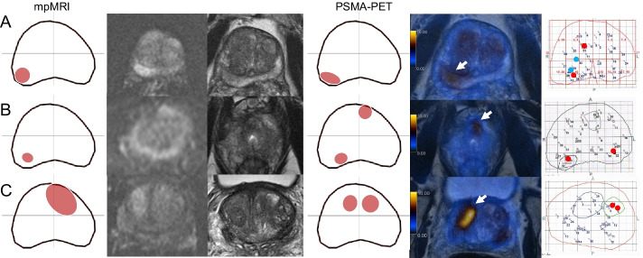
Background: Magnetic resonance imaging (MRI) is recommended by the European Urology Association guidelines as the standard modality for imaging-guided biopsy. Recently positron emission tomography with prostate-specific membrane antigen (PSMA PET) has shown promising results as a tool for this purpose. The aim of this study was to compare the accuracy of positron emission tomography with prostate-specific membrane antigen/magnetic resonance imaging (PET/MRI) using the gallium-labeled prostate-specific membrane antigen (68Ga-PSMA-11) and multiparametric MRI (mpMRI) for pre-biopsy tumour localization and interreader agreement for visual and semiquantitative analysis. Semiquantitative parameters included apparent diffusion coefficient (ADC) and maximum lesion diameter for mpMRI and standardized uptake value (SUVmax) and PSMA-positive volume (PSMAvol) for PSMA PET/MRI.

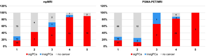
Results: Sensitivity and specificity were 61.4% and 92.9% for mpMRI and 66.7% and 92.9% for PSMA PET/MRI for reader one, respectively. RPE was available in 23 patients and 41 of 47 quadrants with discrepant findings. Based on RPE results, the specificity for both imaging modalities increased to 98% and 99%, and the sensitivity improved to 63.9% and 72.1% for mpMRI and PSMA PET/MRI, respectively. Both modalities yielded a substantial interreader agreement for primary tumour localization (mpMRI kappa = 0.65 (0.52-0.79), PSMA PET/MRI kappa = 0.73 (0.61-0.84)). ICC for SUVmax, PSMAvol and lesion diameter were almost perfect (≥ 0.90) while for ADC it was only moderate (ICC = 0.54 (0.04-0.78)). ADC and lesion diameter did not correlate significantly with Gleason score (ρ = 0.26 and ρ = 0.16) while SUVmax and PSMAvol did (ρ = - 0.474 and ρ = - 0.468).

Conclusions: PSMA PET/MRI has similar accuracy and reliability to mpMRI regarding primary prostate cancer (PCa) localization. In our cohort, semiquantitative parameters from PSMA PET/MRI correlated with tumour grade and were more reliable than the ones from mpMRI.

Keywords: ADC; Biopsy guidance; Interreader agreement; PET/MRI; PSMA PET; Primary staging; SUVmax; Targeted biopsy; Template biopsy; mpMRI.


Conflict of interest statement

IAB has received research grants and speaker honorarium from GE Healthcare, research grants from Swiss Life, and speaker honorarium from Bayer Health Care and Astellas Pharma AG. MM received speaker fees from GE Healthcare. The Department of Nuclear Medicine holds an institutional Research Contract with GE Healthcare. NJR has provided consultancy services (advisory board member) to F. Hoffmann- La Roche AG. ASB received research grants from the Prof. Dr. Max Cloëtta Foundation, medAlumni UZH, and the Swiss Society of Radiology. All other authors declare no competing interests.

Figures

Fig. 1
Fig. 1
Patient selection. After signing the informed consent, three patients refused PSMA PET/MRI and other four patients gave up participation before the biopsy. mpMRI images from three patients were not available for review
Fig. 2
Fig. 2
Example of the method used for lesion localization. A Readout of PSMA PET/MRI by one of the readers with the prostate gland divided in 4 quadrants (Q1: anterior right; Q2: anterior left; Q3: posterior left and Q4: posterior right). The reader delineated three areas of 68Ga-PSMA-11 uptake and labeled area 1 as suspicious for malignancy and areas 2 and 3 as benign/physiological. B 68Ga-PSMA-11 PET/MRI images of this patient show physiological bilateral uptake in the central zone (arrows) and a suspicious area with intense uptake in the posterior peripheral zone on the left (D). C histopathological map automatically generated by the biopsy fusion software with numbered biopsy cores, red spots represent the localization of needles with Gleason score ≥ 3 + 4, confirming the suspicious lesion on Q3 and showing another lesion on Q1 not depicted on PSMA PET/MRI. For the analysis , Q1 was considered false-negative, Q2 true-negative, Q3 true-positive and Q4 false-negative (lesion crossing the midline not depicted by imaging)
Fig. 3
Fig. 3
Imaging findings in relation to biopsy results. Quadrant-based (n = 156) biopsy results in relation to PIRADS on mpMRI and to an adaptation of the same scale for focal uptake on PSMA PET/MRI (1 = no focal uptake; 2 = benign; 3 = undetermined; 4 = suspicious for malignancy ≤ 1.5 cm; 5 = suspicious for malignancy > 1.5 cm). csPCa = clinically significant cancer (red, GS ≥ 3 + 4); ciPCa = clinically insignificant cancer (blue, GS 3 + 3)
Fig. 4
Fig. 4
Imaging and histopathological findings of cases with discordance between PSMA PET/MRI and mpMRI findings. Each line corresponds to one patient. From left to right: mpMRI readout, DWI, T2-w, PSMA PET/MRI readout, fusion PSMA PET/MRI and template biopsy map. Readouts:.lesions in red were classified by the readers as suspicious while the ones in green were classified as non-suspicious. Template biopsy maps: red dots correspond to GS ≥ 3 + 4 biopsy cores and blue dots to GS 3 + 3. A The lesion in the left posterior quadrant was depicted on both mpMRI and PSMA PET, corresponding to csPCa on template biopsy, but the two lesions in the right quadrants were only seen on PSMA PET (arrow in the anterior one). B PSMA PET and mpMRI were concordant regarding the lesions in the anterior right and posterior left quadrants but the apex lesion crossing the midline to the posterior right quadrant was only seen on PSMA PET (arrow). C The lesion in the right posterior quadrant was seen on mpMRI but not on PSMA PET because physiological uptake in the central zones impaired the visual analysis
Fig. 5
Fig. 5
Imaging and histopathological findings of cases in which imaging findings of both mpMRI and PSMA PET were false-positive or false-negative using template biopsy as reference standard. Each line corresponds to one patient. From left to right: mpMRI readout, DWI, T2-w, PSMA PET/MRI readout, fusion PSMA PET/MRI and template biopsy map. A Both imaging modalities depicted the lesion in the posterior right quadrant (arrows) but missed the lesion in the anterior right one (Pat. 7). B Both imaging modalities depicted the lesion in the posterior right quadrant (not shown) but missed the left one and PSMA PET was also false-positive for the anterior left quadrant (arrow). C Imaging was false-positive in the anterior right quadrant, on mpMRI the lesion seems to cross the midline while in PSMA PET the uptake suggests a second lesion (arrow)

References

    1. Ahdoot M, et al. MRI-targeted, systematic, and combined biopsy for prostate cancer diagnosis. N Engl J Med. 2020;382(10):917–928. doi: 10.1056/NEJMoa1910038. - DOI - PMC - PubMed
    1. Ahmed HU, et al. Diagnostic accuracy of multi-parametric MRI and TRUS biopsy in prostate cancer (PROMIS): a paired validating confirmatory study. Lancet. 2017;389(10071):815–822. doi: 10.1016/S0140-6736(16)32401-1. - DOI - PubMed
    1. American Joint Committee on Cancer. Amin MB. AJCC cancer staging manual. New York: Springer; 2017.
    1. Basha MAA, et al. (68)Ga-PSMA-11 PET/CT in newly diagnosed prostate cancer: diagnostic sensitivity and interobserver agreement. Abdom Radiol (NY) 2019;44(7):2545–2556. doi: 10.1007/s00261-019-02006-2. - DOI - PubMed
    1. Bodar YJL, et al. Detection of prostate cancer with (18)F-DCFPyL PET/CT compared to final histopathology of radical prostatectomy specimens: is PSMA-targeted biopsy feasible? The DeTeCT trial. World J Urol. 2020;39:2439–2446. doi: 10.1007/s00345-020-03490-8. - DOI - PMC - PubMed

LinkOut - more resources