

doi: 10.1038/s41392-022-01031-w.
Spatial control of robust transgene expression in mouse artery endothelium under ultrasound guidance
Affiliations
- PMID: 35843985
- PMCID: PMC9288995
- DOI: 10.1038/s41392-022-01031-w
Item in Clipboard
Spatial control of robust transgene expression in mouse artery endothelium under ultrasound guidance
Signal Transduct Target Ther.
.
No abstract available
Conflict of interest statement
The authors declare no competing interests.
Figures
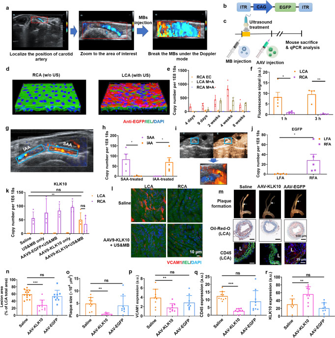
Spatial control of transgene expression in mouse artery with ultrasound. a The mouse carotid artery can be localized by color Doppler ultrasound imaging. By changing the size and position of color box, the ultrasound image can be zoomed to the area of interest so that ultrasound was focused to this area. After MBs injection, the MBs can be destroyed, as evidenced by the color “blooming” phenomenon. b AAV gene vector carrying the expression cassette for EGFP. ITR, internal terminal repeats; CAG, a hybrid construct consisting of the cytomegalovirus (CMV) enhancer fused to the chicken beta-actin promoter. c Illustration of UMGAAV protocol. After MB injection, ultrasound irradiation was applied to the area of interest for 30 s. Following AAV injection, the mouse was sacrificed at specific time points for qPCR analysis using 18S as an internal control. d Representative 3-D view of confocal assessment of EGFP expression in LCA and RCA. EGFP was immunostained with EGFP antibody (red). The DAPI staining marks cell nuclei (blue). The green autofluorescence was from the internal elastic lamina (IEL). e Transgene expression of EGFP in endothelial cells (ECs) and medial and adventitial layer (M + A) of LCA and RCA at different time points post AAV injection. The time interval between MB and AAV injection was ~5 min. f Quantification of AAV fluorescence in LCA and RCA. Fluorescently-labeled AAV was injected after ultrasound treatment in LCA and the mice were sacrificed at 1 h and 3 h post injection. The AAV fluorescence in the arteries were quantified. g Representative image of mouse suprarenal abdominal aorta (SAA) and infrarenal abdominal aorta (IAA) with color Doppler imaging. h Transgene expression of EGFP in SAA and IAA when SAA or IAA was treated with ultrasound. i The mouse femoral artery can be detected with contrast-enhanced ultrasound imaging. After focusing the ultrasound to the major branch of femoral artery, the MBs can be selectively destroyed with color Doppler ultrasound imaging. j Transgene expression of EGFP in mouse femoral artery when right femoral artery (RFA) was treated while the left femoral artery (LFA) was untreated. k Expression of KLK10 in arterial endothelium in mice with different treatments. Saline, mice were injected with saline only; US&MB only, LCA were treated with ultrasound after MB injection; AAV9-EGFP + US&MB, the mice were injected with AAV9 expressing EGFP after ultrasound treatment; AAV9-KLK10 only, the mice were injected with AAV9-KLK10 without ultrasound treatment; AAV9-KLK10 + US&MB, the mice were injected with AAV9-KLK10 after ultrasound treatment. l
En face VCAM1 staining of carotid artery. The arteries were immunostained with VCAM1 antibody (red). The DAPI staining marks cell nuclei (blue). The green autofluorescence was from IEL. m Representative bright-field images of aortic trees (top lane) and the Oil-Red-O staining (middle lane) and anti-CD45 immunostaining (bottom lane) of frozen sections prepared from the middle parts of these arteries. CD45 was immunostained in red. The DAPI staining marks cell nuclei (blue). The green autofluorescence was from IEL. L, lumen of the artery. n Quantification of lesion area in LCA. o Quantification of plaque size determined with the Oil-Red-O-stained sections. p–r Quantification of VCAM1 (p), CD45 (q), and KLK10 (r) expression according to the immunostaining of the frozen sections. Data shown as mean ± s.e.m; *P < 0.05; **P < 0.01; ***P < 0.001; ns, P > 0.05 as determined by Student’s t-test
References
Publication types
MeSH terms
Grants and funding
LinkOut - more resources
Full Text Sources
