. 2022 Dec;530(17):2977-2993.
doi: 10.1002/cne.25389. Epub 2022 Jul 17.

Olfactory placode generates a diverse population of neurons expressing GnRH, somatostatin mRNA, neuropeptide Y, or calbindin in the chick forebrain

Affiliations

Olfactory placode generates a diverse population of neurons expressing GnRH, somatostatin mRNA, neuropeptide Y, or calbindin in the chick forebrain

Shizuko Murakami et al. J Comp Neurol. 2022 Dec.

Abstract

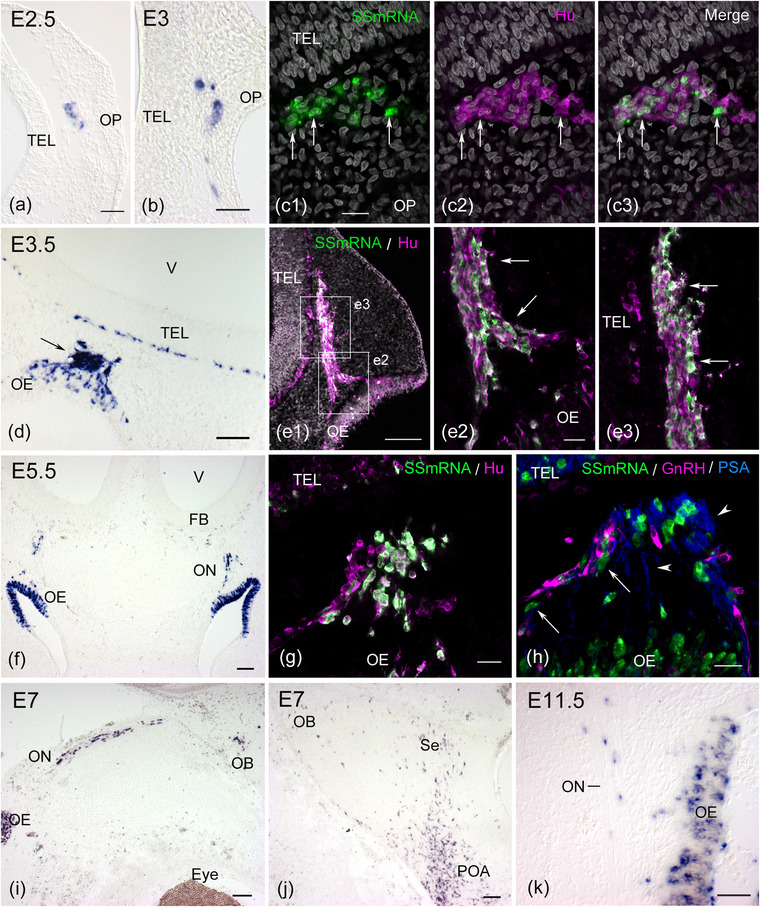
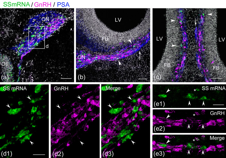
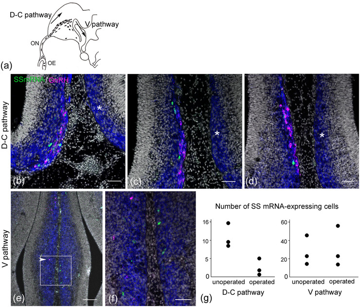
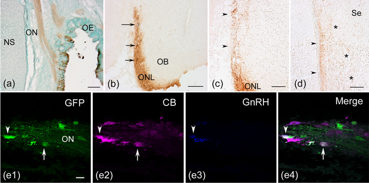
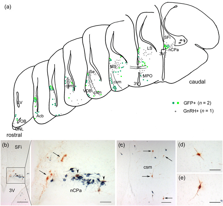
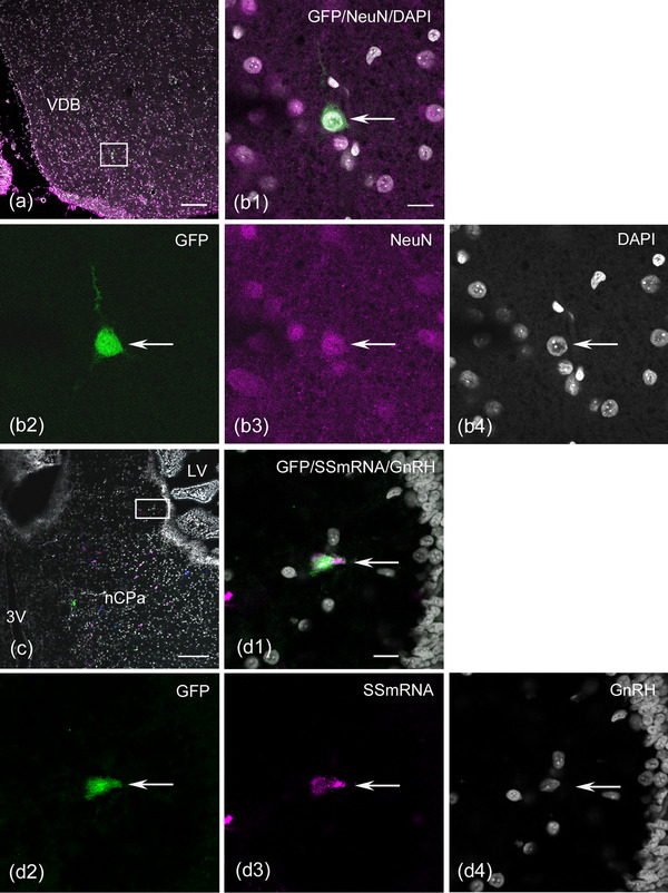
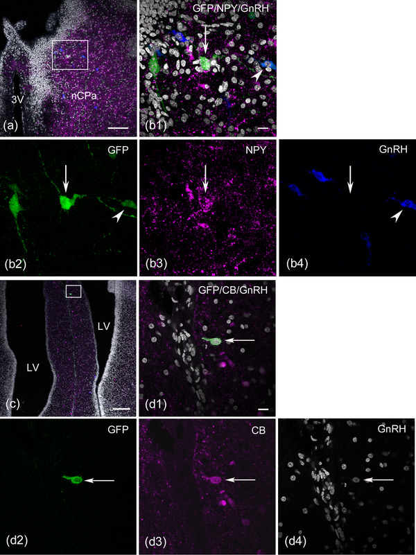
The olfactory placode (OP) of vertebrates generates several classes of migrating cells, including hypothalamic gonadotropin-releasing hormone (GnRH)-producing neurons, which play essential roles in the reproduction system. Previous studies using OP cell labeling have demonstrated that OP-derived non-GnRH cells enter the developing forebrain; however, their final fates and phenotypes are less well understood. In chick embryos, a subpopulation of migratory cells from the OP that is distinct from GnRH neurons transiently expresses somatostatin (SS). We postulated that these cells are destined to develop into brain neurons. In this study, we examined the expression pattern of SS mRNA in the olfactory-forebrain region during development, as well as the destination of OP-derived migratory cells, including SS mRNA-expressing cells. Utilizing the Tol2 genomic integration system to induce long-term fluorescent protein expression in OP cells, we found that OP-derived migratory cells labeled at embryonic day (E) 3 resided in the olfactory nerve and medial forebrain at E17-19. A subpopulation of green fluorescent protein (GFP)-labeled GnRH neurons that remained in the olfactory nerve was considered to comprise terminal nerve neurons. In the forebrain, GFP-labeled cells showed a distribution pattern similar to that of GnRH neurons. A large proportion of GFP-labeled cells expressed the mature neuronal marker NeuN. Among the GFP-labeled cells, the percentage of GnRH neurons was low, while the remaining GnRH-negative neurons either expressed SS mRNA, neuropeptide Y, or calbindin D-28k or did not express any of them. These results indicate that a diverse population of OP-derived neuronal cells, other than GnRH neurons, integrates into the chick medial forebrain.

Keywords: GnRH neurons; Tol2-transposon; forebrain; neuronal migration; olfactory placode; somatostatin mRNA; terminal nerve.


Conflict of interest statement

The authors declare no conflict of interest.

Figures

FIGURE 1
FIGURE 1
Somatostatin (SS) mRNA expression in the developing chick olfactory‐forebrain region. (a) Cross section through the head of an E2.5 (HH16) chick embryo. Few SS mRNA‐positive cells are seen in the mescenchymal tissue beneath the olfactory placode (OP). (b) At E3 (HH17), SS mRNA‐expressing cells are found in the epithelium of the OP and mesenchymal tissue. (c) In situ hybridization on a cross section followed by immunostaining for HuC/D labels a cluster of migratory cells from the OP at HH17. SS mRNA‐expressing cells that coexpress HuC/D are found in the HuC/D‐positive cellular mass‐like structure (arrows in c1–c3). (d) Cross section through the olfactory‐forebrain region of an E3.5 (HH21) chick embryo. A cluster of migratory cells from the olfactory epithelium (OE) strongly expresses SS mRNA (arrow). (e) Sagittal section through the head of an E3.5 (HH19) embryo. HuC/D‐positive migratory cells form the cellular cord between the OE and telencephalon (TEL). Many SS mRNA‐expressing cells are found in the cellular cord (e1). Regions similar to those outlined by the squares show that SS mRNA and HuC/D double‐positive cells are located in a thick cellular cord (arrows in e2 and e3). (f–h) Cross sections through the olfactory‐forebrain region of E5.5 chick embryos. Intense expression of SS mRNA is seen in the OE and migratory cells along the olfactory nerve (ON) (f). SS mRNA‐expressing cells that coexpress HuC/D migrate from the OE (g). Some SS mRNA‐expressing cells comigrated with gonadotropin‐releasing hormone (GnRH) neurons (arrows in h). These migratory cells are associated with olfactory fibers that express highly polysialylated neural cell adhesion molecule (PSA‐NCAM, arrowheads). (i–j) Sagittal section through the head of an E7 chick embryo. SS mRNA‐positive cells are seen in the OE, ON, and olfactory bulb (OB). SS mRNA‐expressing cells are sparsely distributed in the dorsal forebrain, including the septum region (Se), while many SS mRNA‐expressing cells are found in the preoptic area (POA) of the ventral forebrain. (k) Sagittal section through the olfactory region of an E11.5 chick embryo. SS mRNA is still expressed in the OE and the cells associated with the ON. (a), (b), (d), and (k): Viewed with Nomarski optics. FB, forebrain. Scale bars: 20 μm in c1, e2, g, and h; 50 μm in a, b, and k; 100 μm in d, e1, f, i, and j
FIGURE 2
FIGURE 2
Somatostatin (SS) mRNA‐expressing cells are accompanied by gonadotropin‐releasing hormone (GnRH) neurons in the olfactory‐forebrain region. Coronal sections through the olfactory‐forebrain region of E6.5−7 embryos. (a) SS mRNA‐expressing cells (green) and GnRH neurons (magenta) migrate along the medial portion of the olfactory nerve (ON, blue) that express highly polysialylated neural cell adhesion molecule (PSA‐NCAM). (b) A small subset of PSA‐NCAM‐immunoreactive (‐ir) fibers branch medially from the ON and extend to the medial forebrain (arrow). SS mRNA‐expressing cells and GnRH neurons migrate along these fibers into the medial forebrain (arrowheads). (c) At a more caudal level of panel (b), a small number of SS mRNA‐expressing cells are seen in the parenchyma of the medial forebrain (arrowheads), where migrating GnRH neurons are aligned continuously along the PSA‐NCAM‐ir fibers. A small fascicle of the medial branch of the ON is seen to extend along the medial forebrain (arrow). (d) A confocal stacked image of an olfactory region of an E6.5 chick embryo shows that SS mRNA‐expressing cells are immunonegative for GnRH (arrowheads in d1–d3). (e) A confocal stacked image of the medial forebrain of an E7 chick embryo shows that almost none of the SS mRNA‐expressing cells express GnRH. In this region, three SS mRNA‐expressing cells adjacent to GnRH neurons form a distinct cell population from GnRH neurons (arrowheads in e1–e3), while only one SS mRNA‐expressing cell exhibits very weak GnRH immunoreactivity (asterisk in e1–e3). Scale bars: 50 μm in a, b, and c; 20 μm in d1 and e1. Abbreviations: FB, forebrain; LV, lateral ventricle; OE, olfactory epithelium
FIGURE 3
FIGURE 3
Unilateral olfactory placodectomy induces a loss of somatostatin (SS) mRNA‐expressing cells and gonadotropin‐releasing hormone (GnRH) neurons in the migratory course of the forebrain. Coronal section through the forebrain of an E7.5 embryo after unilateral placodectomy at E3.5. (a) Schematic figure shows the proposed migration pathway of GnRH neurons in the forebrain (Murakami et al., 2010). (b–d) Coronal sections through the forebrain are aligned rostrally (b) to caudally (d) at different intervals. On the unoperated side, a small number of SS mRNA‐expressing cells (green) migrate in association with GnRH neurons (magenta) into the medial forebrain (b). Highly polysialylated neural cell adhesion molecule (PSA‐NCAM) immunoreactivity (blue) highlights immature neurons in the developing forebrain. SS mRNA‐expressing cells are found within a cluster of migratory GnRH neurons in the medial forebrain surface (c–d). On the operated side, no SS mRNA‐expressing cells or GnRH neurons are seen in the equivalent region on the unoperated side (asterisk in b–d). (e–f) At the level of the V pathway of GnRH neurons, scattered GnRH neurons migrate ventrally on the unoperated side (arrowhead in e) but not on the operated side. An enlarged image of panel (e) demonstrates that SS mRNA‐expressing cells are detected in the V pathway of GnRH neurons on both sides (f). (g) Quantification of the number of SS mRNA‐expressing cells in the D‐C and V pathways of GnRH neurons (n = 3 embryos), which were counted in every fifth coronal section. The D‐C pathway is characterized by a small cluster of GnRH neurons that are aligned along the medial forebrain surface in coronal sections. Thus, the counting of SS mRNA‐expressing cells in the D‐C pathway was performed within this restricted region. Ablation of the olfactory placode induces a remarkable reduction in the number of SS mRNA‐expressing cells in the D‐C pathway on the operated side. Scale bars, 50 μm in b, c, d, and f; 100 μm in e. Abbreviations: OE, olfactory epithelium; ON, olfactory nerve
FIGURE 4
FIGURE 4
Persistent green fluorescent protein (GFP) expression in the peripheral olfactory region and neuronal phenotypes of GFP‐labeled cells. The Tol2‐mediated GFP construct was electroporated into the olfactory placode at HH18 (E3), and horizontal (a and e) or coronal (b–d) sections were obtained at E17−19. (a) A part of the olfactory epithelium (OE) and the olfactory nerve (ON) are labeled by transfer to the Tol2‐GFP construct. (b) The GFP‐labeled ON fibers terminate in the olfactory nerve layer (ONL) of the olfactory bulb (OB). GFP‐labeled cells (short arrows) and a gonadotropin‐releasing hormone (GnRH) neuron (long arrow) are seen in the GFP‐positive ONL. (c) A small number of GFP‐labeled fibers apart from the ON extend to the medial forebrain surface caudally (arrowheads). (d) A small fascicle of GFP‐labeled fibers is seen to terminate in the medial edge of the dorsocaudal portion of the septum (arrowheads). Some GFP‐labeled fibers are seen to disperse into the deep layer of the septum (asterisks). (e) A cluster of GFP‐labeled cells is observed in the ON, and two labeled cells coexpress calbindin D‐28k (CB). One GFP‐labeled cell coexpresses CB but not GnRH (arrow in e1, e2, and e4), while another GFP‐labeled cell coexpresses both CB and GnRH (arrowhead in e1–e4). (c) and (d): Viewed with Nomarski optics. Scale bars: 100 μm in a and b; 50 μm in c and d; 20 μm in e. Abbreviations: NS, nasal septum; Se, septum
FIGURE 5
FIGURE 5
Distribution patterns of green fluorescent protein (GFP)‐labeled cells and gonadotropin‐releasing hormone (GnRH) neurons in the forebrain of E17−19 embryos. Coronal sections through the forebrain electroporated with the Tol2‐mediated GFP plasmid into the olfactory placode at HH18 (E3) and processed at E17−19. (a) Schematic drawings of hemisections through the forebrain of E18 chick embryos showing the distribution of GFP‐labeled cells and GnRH neurons. They are arranged from rostral to caudal at 80−400 μm intervals. The location of GFP‐labeled cells is represented by the data of two embryos (green and yellow‒green circles, 94 cells), while that of GnRH neurons is the data of one embryo (gray circles, 207 neurons). The distribution pattern was largely similar between the two cell groups. GFP‐labeled cells that coexpressed GnRH were excluded. (b) GFP‐labeled cells are seen in and around the bed nucleus of the pallial commissure (nCPa) at the dorsocaudal level of the septum, including the septofimbrial nucleus (SFi). GFP‐labeled GnRH‐negative cells are found near the nCPa (arrows). A cluster of GnRH neurons is seen in the nCPa, where two GnRH neurons coexpress GFP (arrowheads). (c) GFP‐labeled cells are located in the vertical limb of the diagonal band (VDB) at the ventral part of the subpallium (arrows). (d and e) GFP‐labeled cells have multipolar processes. Scale bars: 200 μm in b; 100 μm in c; 50 μm in the large panels of b, d, and e. Abbreviations: ac, anterior commissure; Acb, accumbens nucleus; csm, corticoseptomesencephalic tract; LS, lateral septum; MPO, medial preoptic area; MS, medial septum; OB, olfactory bulb; ONL, olfactory nerve layer; Se, septum; 3V, third ventricle
FIGURE 6
FIGURE 6
Green fluorescent protein (GFP)‐labeled olfactory placode (OP)‐derived cells coexpress neuronal nuclei (NeuN) or somatostatin (SS) mRNA in the medial forebrain. Coronal sections through the forebrain of E17 or E19 embryos electroporated with the Tlo2‐mediated EGFP plasmid into the OP at HH18 (E3). (a and b) Confocal single image at the ventral portion of the rostral‐medial forebrain. An enlarged image of panel (a) demonstrates that a GFP‐labeled cell expresses NeuN (arrow in b1–b4). (c and d) The combination of fluorescent in situ hybridization and immunohistochemistry reveals that GFP‐labeled cells coexpress SS mRNA in the bed nucleus of the pallial commissure (nCPa). Pseudocolored confocal images for GFP and SS mRNA were produced using Zeiss ZEN. GFP‐labeled cells (green), SS mRNA‐expressing cells (magenta), and gonadotropin‐releasing hormone (GnRH) neurons (blue) are seen in the nCPa (c). An enlarged confocal single image of panel (c) shows a GFP‐labeled cell expressing SS mRNA negative for GnRH (arrow in d1–d4). Scale bars: 100 μm in a and c; 10 μm in b1 and d1. Abbreviations: LV, lateral ventricle; 3V, third ventricle; VDB, vertical limb of the diagonal band
FIGURE 7
FIGURE 7
Green fluorescent protein (GFP)‐labeled olfactory placode‐derived cells also coexpress neuropeptide Y (NPY) or calbindin D‐28k (CB) in the medial forebrain at E17 or E19. (a and b) Confocal single image at the dorsocaudal level of the septum region, including the bed nucleus of the pallial commissure (nCPa). An enlarged image of panel (a) shows the colocalization of NPY in the GFP‐labeled cell. The stacked image (z‐projection image stacks) reveals that many GFP‐labeled cells are observed in the nCPa (b1 and b2). A GFP‐labeled cell expresses NPY (arrow in b1‐ b3) but does not express gonadotropin‐releasing hormone (GnRH) (arrow in b4), while another labeled cell expresses GnRH (arrowhead in b1, b2, and b4), and the remaining labeled cell does not express either NPY or GnRH. (c and d) Confocal single image of triple immunostaining with GFP, CB, and GnRH antibodies shows a GFP‐labeled CB‐expressing neuron in the rostral medial forebrain caudal to the olfactory bulb. An enlarged image of panel (c) demonstrates that a GFP‐labeled cell expresses CB (arrow in d1‐ d3) but does not express GnRH (arrow in d4). Scale bars: 100 μm in a and c; 10 μm in b1 and d1. Abbreviations: LV, lateral ventricle; 3V, third ventricle

Similar articles

Cited by

References

    1. Aguillon, R. , Batut, J. , Subramanian, A. , Madelaine, R. , Dufourcq, P. , Schilling, T. F. , & Blader, P. (2018). Cell‐type heterogeneity in the early zebrafish olfactory epithelium is generated from progenitors within preplacodal ectoderm. eLife, 7, e32041. - PMC - PubMed
    1. Akutsu, S. , Takada, M. , Ohki‐Hamazaki, H. , Murakami, S. , & Arai, Y. (1992). Origin of luteinizing hormone‐releasing hormone (LHRH) neurons in the chick embryo: Effect of the olfactory placode ablation. Neuroscience Letters, 142, 241–244. - PubMed
    1. Airaksinen, M. S. , Eilers, J. , Garaschuk, O. , Thoenen, H. , Konnerth, A. , & Meyer, M. (1997). Ataxia and altered dendritic calcium signaling in mice carrying a targeted null mutation of the calbindin D28k gene. Proceedings of the National Academy of Sciences of the United State of America, 94, 1488–1493. - PMC - PubMed
    1. Baimbridge, K. G. , Celio, M. R. , & Rogers, J. H. (1992). Calcium‐binding proteins in the nervous system. Trends in Neuroscience, 15, 303–308. - PubMed
    1. Blähser, S. (1984). Peptidergic pathways in the avian brain. The Journal of Experimental Zoology, 232, 397–403. - PubMed

Publication types