

Hypophysial angiogenesis decodes annual time and underlies physiological adaptation to seasonal changes in the environment
- PMID: 35844178
- PMCID: PMC9796326
- DOI: 10.1002/jez.2639
Hypophysial angiogenesis decodes annual time and underlies physiological adaptation to seasonal changes in the environment
Abstract
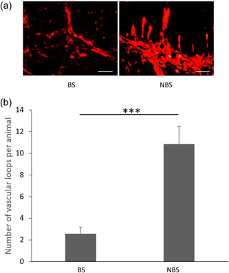
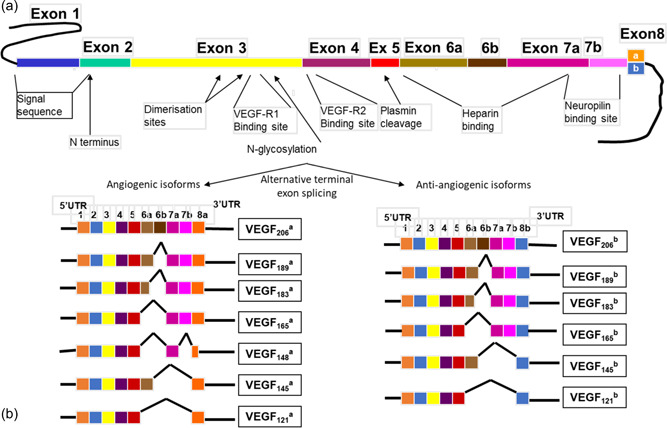
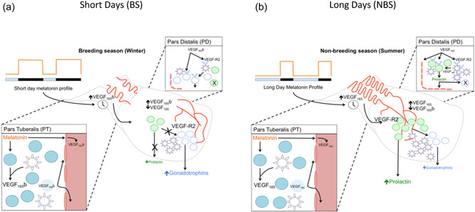
Adaptation to annual changes in the environment is controlled by hypophysial hormones. In temperate zones, photoperiod is the primary external cue that regulates annual biological cycles and is translated by the pattern of melatonin secretion acting primarily in the hypophysial pars tuberalis. Angiogenic mechanisms within this tissue contribute to decode the melatonin signal through alternative splicing of the vascular endothelial growth factor A (VEGF-A) gene in both the pars tuberalis and the capillary loops of the infundibulum. The resulting melatonin-evoked differential productions of VEGF-A isoforms will induce seasonal remodeling of the vascular connection between the hypothalamus and hypophysis, and act as paracrine messengers in the pars distalis to generate the required seasonal endocrine response. Specifically, the long melatonin signal in winter upregulates antiangiogenic VEGF-A isoforms, which will reduce the number of vascular loops and the density of VEGF receptors in endocrine and folliculo-stellate (FS) cells, inhibit prolactin secretion, and stimulate FSH. In contrast, the short melatonin signal in summer upregulates proangiogenic VEGF-A isoforms that will increase the number of vascular loops and the density of VEGF receptors in endocrine and FS cells, stimulate prolactin secretion, and suppress FSH. A similar system has been identified in long day seasonal breeders, revealing that this is a conserved mechanism of adaptation across species. Thus, an angiogenesis-based, intrahypophysial system for annual time measurement controls local microvascular plasticity and conveys the photoperiodic signal readout from the melatonin sensitive pars tuberalis to the endocrine cells of the pars distalis to regulate seasonal adaptation to the environment.
Keywords: VEGE-A; angiogenesis; melatonin; pars tuberalis; photoperiod; pituitary gland; prolactin.
© 2022 The Authors. Journal of Experimental Zoology Part A: Ecological Genetics and Physiology published by Wiley Periodicals LLC.
Conflict of interest statement
The author declares no conflict of interest.
Figures
References
-
- Acosta, M. & Mohamed, F. (2009). Pituitary pars intermedia of male viscacha (Lagostomus maximus maximus): A morphometric study of seasonal and age‐related changes in immunohistochemistry. Cells Tissues Organs, 190, 219–229. - PubMed
-
- Acosta, M. & Mohamed, F. (2011). Effect of the photoperiod and administration of melatonin on folliculostellate cells of the pituitary pars distalis of adult male viscacha (Lagostomus maximus maximus). Acta Histochemica, 113, 640–646. - PubMed
-
- Allaerts, W. , Engelborghs, Y. , Vanoostveldt, P. , & Denef, C. (1990). Evidence that folliculostellate cells do not impede the permeability of intercellular spaces to molecular diffusion in three‐dimensinal aggregate cell cultures of the rat anterior pituitary. Endocrinology, 127, 1517–1525. - PubMed
-
- Allaerts, W. & Vankelecom, H. (2005). History and perspectives of pituitary folliculo‐stellate cell research. European Journal of Endocrinology, 153, 1–12. - PubMed
Publication types
MeSH terms
Substances
Grants and funding
LinkOut - more resources
Full Text Sources
Miscellaneous
