Abnormal Voxel-Based Degree Centrality in Patients With Postpartum Depression: A Resting-State Functional Magnetic Resonance Imaging Study
- PMID: 35844214
- PMCID: PMC9280356
- DOI: 10.3389/fnins.2022.914894
Abnormal Voxel-Based Degree Centrality in Patients With Postpartum Depression: A Resting-State Functional Magnetic Resonance Imaging Study
Abstract
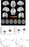
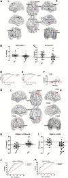
Postpartum depression (PPD) is a major public health concern with significant consequences for mothers, their children, and their families. However, less is known about its underlying neuropathological mechanisms. The voxel-based degree centrality (DC) analysis approach provides a new perspective for exploring the intrinsic dysconnectivity pattern of whole-brain functional networks of PPD. Twenty-nine patients with PPD and thirty healthy postpartum women were enrolled and received resting-state functional magnetic resonance imaging (fMRI) scans in the fourth week after delivery. DC image, clinical symptom correlation, and seed-based functional connectivity (FC) analyses were performed to reveal the abnormalities of the whole-brain functional network in PPD. Compared with healthy controls (HCs), patients with PPD exhibited significantly increased DC in the right hippocampus (HIP.R) and left inferior frontal orbital gyrus (ORBinf.L). The receiver operating characteristic (ROC) curve analysis showed that the area under the curve (AUC) of the above two brain regions is all over 0.7. In the seed-based FC analyses, the PPD showed significantly decreased FC between the HIP.R and right middle frontal gyrus (MFG.R), between the HIP.R and left median cingulate and paracingulate gyri (DCG.L), and between the ORBinf.L and the left fusiform (FFG.L) compared with HCs. The PPD showed significantly increased FC between the ORBinf.L and the right superior frontal gyrus, medial (SFGmed.R) compared with HCs. Mean FC between the HIP.R and DCG.L positively correlated with EDPS scores in the PPD group. This study provided evidence of aberrant DC and FC within brain regions in patients with PPD, which was associated with the default mode network (DMN) and limbic system (LIN). Identification of these above-altered brain areas may help physicians to better understand neural circuitry dysfunction in PPD.
Keywords: fMRI; postpartum depression; receiver operating characteristic (ROC) curve analysis; seed-based functional connectivity; voxel-based degree centrality.
Copyright © 2022 Zhang, Li, Liu, Hou and Zhang.
Conflict of interest statement
The authors declare that the research was conducted in the absence of any commercial or financial relationships that could be construed as a potential conflict of interest.
Figures


Similar articles
-
Aberrant structural and functional alterations in postpartum depression: a combined voxel-based morphometry and resting-state functional connectivity study.Front Neurosci. 2023 May 25;17:1138561. doi: 10.3389/fnins.2023.1138561. eCollection 2023. Front Neurosci. 2023. PMID: 37304034 Free PMC article.
-
Disruption of Neural Activity and Functional Connectivity in Adolescents With Major Depressive Disorder Who Engage in Non-suicidal Self-Injury: A Resting-State fMRI Study.Front Psychiatry. 2021 Jun 1;12:571532. doi: 10.3389/fpsyt.2021.571532. eCollection 2021. Front Psychiatry. 2021. PMID: 34140897 Free PMC article.
-
Aberrant voxel-based degree centrality in Parkinson's disease patients with mild cognitive impairment.Neurosci Lett. 2021 Jan 10;741:135507. doi: 10.1016/j.neulet.2020.135507. Epub 2020 Nov 17. Neurosci Lett. 2021. PMID: 33217504
-
Alterations in degree centrality and functional connectivity in tension-type headache: a resting-state fMRI study.Brain Imaging Behav. 2024 Aug;18(4):819-829. doi: 10.1007/s11682-024-00875-w. Epub 2024 Mar 21. Brain Imaging Behav. 2024. PMID: 38512647 Review.
-
Functional changes of default mode network and structural alterations of gray matter in patients with irritable bowel syndrome: a meta-analysis of whole-brain studies.Front Neurosci. 2023 Oct 24;17:1236069. doi: 10.3389/fnins.2023.1236069. eCollection 2023. Front Neurosci. 2023. PMID: 37942144 Free PMC article.
Cited by
-
Altered brain network centrality in patients with orbital fracture: A resting‑state functional MRI study.Exp Ther Med. 2023 Oct 17;26(6):552. doi: 10.3892/etm.2023.12251. eCollection 2023 Dec. Exp Ther Med. 2023. PMID: 37941594 Free PMC article.
-
Aberrant structural and functional alterations in postpartum depression: a combined voxel-based morphometry and resting-state functional connectivity study.Front Neurosci. 2023 May 25;17:1138561. doi: 10.3389/fnins.2023.1138561. eCollection 2023. Front Neurosci. 2023. PMID: 37304034 Free PMC article.
-
Distance-related functional reorganization predicts motor outcome in stroke patients.BMC Med. 2024 Jun 18;22(1):247. doi: 10.1186/s12916-024-03435-7. BMC Med. 2024. PMID: 38886774 Free PMC article.
-
Biomarkers of reproductive psychiatric disorders.Br J Psychiatry. 2025 Jun;226(6):352-368. doi: 10.1192/bjp.2025.134. Epub 2025 Jun 20. Br J Psychiatry. 2025. PMID: 40538358 Free PMC article. Review.
-
Aberrant regional neural fluctuations and functional connectivity in insomnia comorbid depression revealed by resting-state functional magnetic resonance imaging.Cogn Neurodyn. 2025 Dec;19(1):8. doi: 10.1007/s11571-024-10206-w. Epub 2025 Jan 6. Cogn Neurodyn. 2025. PMID: 39780909
References
LinkOut - more resources
Full Text Sources

