. 2021 Dec;11(23):11301.
doi: 10.3390/app112311301. Epub 2021 Nov 29.

Fe3O4 Nanoparticles: Structures, Synthesis, Magnetic Properties, Surface Functionalization, and Emerging Applications

Affiliations

Fe3O4 Nanoparticles: Structures, Synthesis, Magnetic Properties, Surface Functionalization, and Emerging Applications

Minh Dang Nguyen et al. Appl Sci (Basel). 2021 Dec.

Abstract

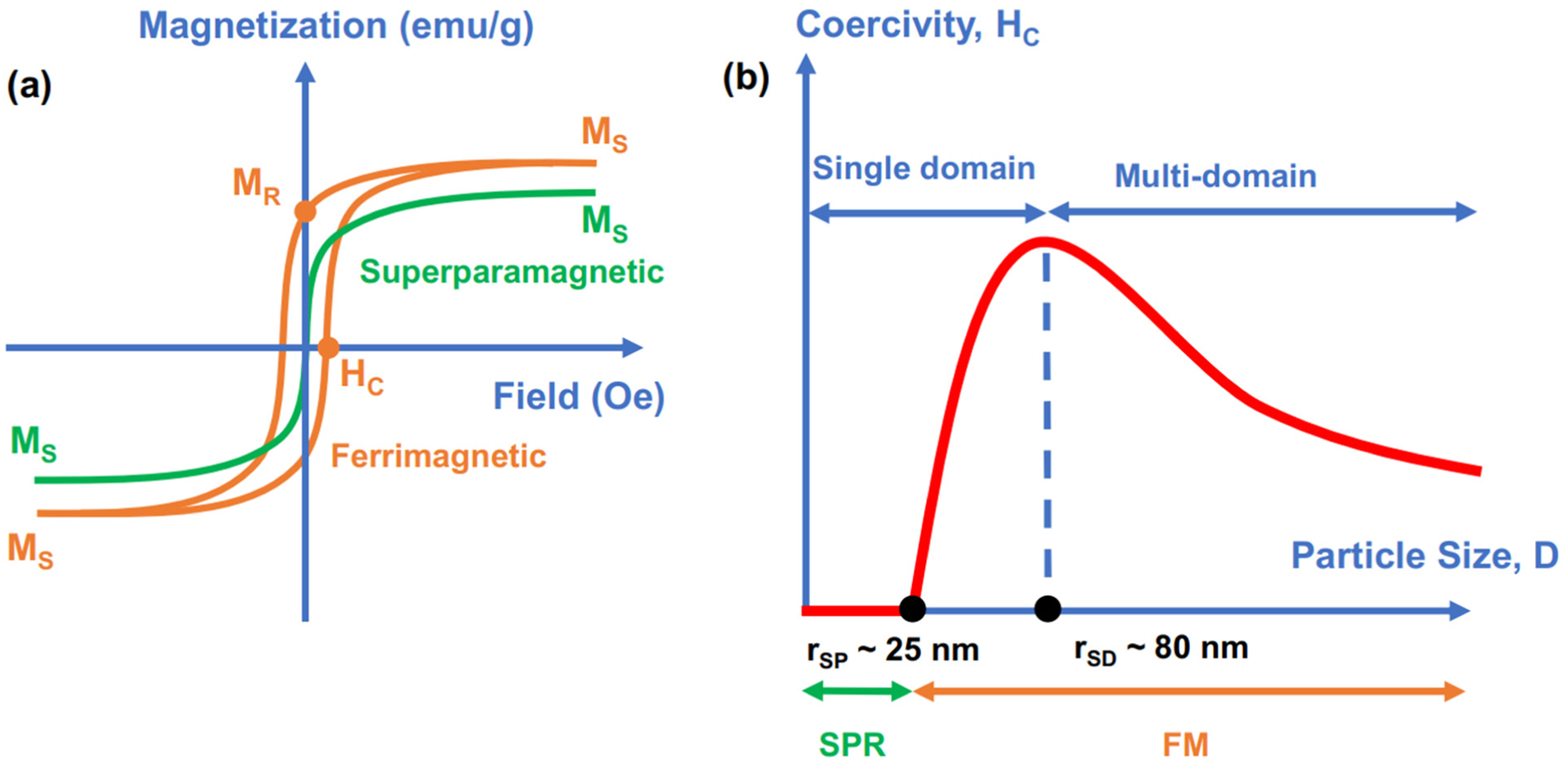
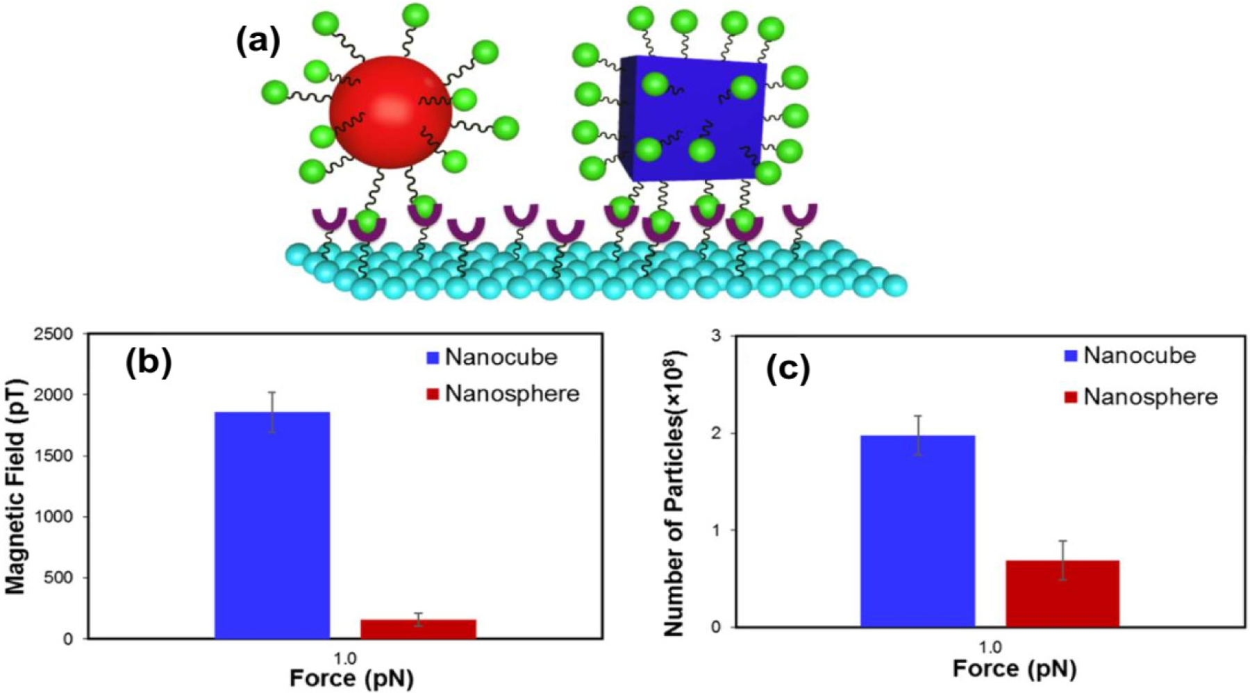
Magnetite (Fe3O4) nanoparticles (NPs) are attractive nanomaterials in the field of material science, chemistry, and physics because of their valuable properties, such as soft ferromagnetism, half-metallicity, and biocompatibility. Various structures of Fe3O4 NPs with different sizes, geometries, and nanoarchitectures have been synthesized, and the related properties have been studied with targets in multiple fields of applications, including biomedical devices, electronic devices, environmental solutions, and energy applications. Tailoring the sizes, geometries, magnetic properties, and functionalities is an important task that determines the performance of Fe3O4 NPs in many applications. Therefore, this review focuses on the crucial aspects of Fe3O4 NPs, including structures, synthesis, magnetic properties, and strategies for functionalization, which jointly determine the application performance of various Fe3O4 NP-based systems. We first summarize the recent advances in the synthesis of magnetite NPs with different sizes, morphologies, and magnetic properties. We also highlight the importance of synthetic factors in controlling the structures and properties of NPs, such as the uniformity of sizes, morphology, surfaces, and magnetic properties. Moreover, emerging applications using Fe3O4 NPs and their functionalized nanostructures are also highlighted with a focus on applications in biomedical technologies, biosensing, environmental remedies for water treatment, and energy storage and conversion devices.

Keywords: Fe3O4 nanoparticles; biomedical applications; biosensing; core-shell structures; energy storage; environmental applications; magnetic properties; nanocomposites; surface functionalization.


Conflict of interest statement

Conflicts of Interest: The authors have no competing financial interest to declare.

Figures

Figure 1.
Figure 1.
Numbers of articles with the keyword “Fe3O4” published in the last 20 years. Data originated from the Web of Science. (Data from 2021 were collected until September 2021).
Figure 2.
Figure 2.
Fe3O4 NPs with various nanostructures and sizes used in emerging biomedical, biosensing, environmental, and energy applications.
Figure 3.
Figure 3.
(a) Magnetization versus applied field (M–H) curves for superparamagnetic (SPM) (green color) and ferrimagnetic (FM) (orange) Fe3O4 nanoparticles and (b) relations between size, coercivity, and magnetic behavior.
Figure 4.
Figure 4.
(a) Schematic synthesis of Fe3O4 NPs with sizes of 7, 8, 9, and 10 nm. Selected TEM images of Fe3O4 NPs with sizes of (b) 7 ± 0.5 nm and (c) 10 ± 0.8 nm. Scale bar 20 nm. Reproduced with permission from ref. [57]. Copyright 2009 American Chemical Society. (d) TEM image of spherical Fe3O4 NPs with a size of 200 nm. Reproduced with permission from ref. [48]. Copyright 2005 John Wiley and Sons.
Figure 5.
Figure 5.
(a) Schematics showing the synthesis of Fe3O4 nanocubes with edge lengths in the 9–80 nm range and (b) the growth mechanism of Fe3O4 nanocubes. Reproduced with permission from ref. [74]. Copyright 2019 American Chemical Society.
Figure 6.
Figure 6.
(a,b) TEM and HR-TEM images of Fe3O4 nanorods. Reproduced with permission from ref. [79]. Copyright 2012 American Chemical Society. (c,d) TEM and HR-TEM images of porous hollow NPs. Reproduced with permission from ref. [87]. Copyright 2009 American Chemical Society. (e) TEM image of 2D hexagonal nanoplates. Reproduced with permission from ref. [85]. Copyright 2010 American Chemical Society. (f,g) TEM images of Fe3O4 tripods and tetrapods. Reproduced with permission from ref. [86]. Copyright 2009 Elsevier.
Figure 7.
Figure 7.
Schematic illustration of the proposed growth model for MNCs. Reproduced with permission from ref. [76]. Copyright 2017 American Chemical Society.
Figure 8.
Figure 8.
(a) TEM image of 22 nm nanocubes encapsulating PEG-phospholipid. (b) Image of colloidal iron oxide nanocubes. In vivo MR images of the tumor site: (c) without colloid injection and (d) after 1 h (intravenous injection). MR contrast effect of ferrimagnetic iron oxide nanocubes with different sizes: (e) T2-weighted MR images obtained with various concentrations of iron in a 3 T field and (f) their color-coded presentation. Reproduced with permission from ref. [106]. Copyright 2012 American Chemical Society.
Figure 9.
Figure 9.
TEM images of colloidal nanoclusters constructed from Fe3O4 nanocube-coated amphiphilic copolymer poly(styrene-comaleic anhydride) to form (a) monomer clusters (1 nanocube), (b) dimers and trimers (2–3 nanocubes), and (c) centrosymmetric clusters (more than 4 nanocubes). (d) Schematic illustration of the preparation of soft colloidal nanoclusters. (e) SAR values for different soft colloidal nanoclusters. Reproduced with permission from ref. [117]. Copyright 2017 American Chemical Society.
Figure 10.
Figure 10.
(a) Synthetic scheme for GOD-encapsulated hollow iron oxide nanoparticles and (b) multifunctional therapeutic strategies for starvation–chemodynamic–hyperthermia using GOD-encapsulated hollow IONPs. Reproduced with permission from ref. [89]. Copyright 2020 American Chemical Society.
Figure 11.
Figure 11.
(a) Magnetic signal of streptavidin-conjugated Fe3O4@SiO2@PAA NPs bound to a biotinylated surface. (b) Schematic illustration of the interaction between magnetic NPs and the surface for protein detection in the EXIRM analysis. (c) EXIRM data for Protein A arising from an exchange between IgG1 and IgG2 subclasses. Reproduced with permission from ref. [43]. Copyright 2018 American Chemical Society.
Figure 12.
Figure 12.
(a) Schematic illustration of the contact area in the biosensing of nanocubic and nanospherical Fe3O4. FIRMS data showing the (b) magnetization profiles versus applied force of nanocubes and nanospheres with similar volumes and (c) number of retained nanoparticles on the sensor at 1 pN. Reproduced with permission from ref. [42]. Copyright 2017 American Chemical Society.

Similar articles

Cited by

References

    1. Jeong U; Teng X; Wang Y; Yang H; Xia Y Superparamagnetic Colloids: Controlled Synthesis and Niche Applications. Adv. Mater 2007, 19, 33–60.
    1. Lu A-H; Salabas EL; Schüth F Magnetic Nanoparticles: Synthesis, Protection, Functionalization, and Application. Angew. Chem. Int. Ed 2007, 46, 1222–1244. - PubMed
    1. Singamaneni S; Bliznyuk VN; Binek C; Tsymbal EY Magnetic Nanoparticles: Recent Advances in Synthesis, Self-Assembly and Applications. J. Mater. Chem 2011, 21, 16819–16845.
    1. Frey NA; Peng S; Cheng K; Sun S Magnetic Nanoparticles: Synthesis, Functionalization, and Applications in Bioimaging and Magnetic Energy Storage. Chem. Soc. Rev 2009, 38, 2532–2542. - PMC - PubMed
    1. Lisjak D; Mertelj A Anisotropic Magnetic Nanoparticles: A Review of Their Properties, Syntheses and Potential Applications. Prog. Mater. Sci 2018, 95, 286–328.

LinkOut - more resources