

Subacute toxic effects of silver nanoparticles oral administration and withdrawal on the structure and function of adult Albino Rats' hepatic tissue
- PMID: 35844407
- PMCID: PMC9280256
- DOI: 10.1016/j.sjbs.2022.02.054
Subacute toxic effects of silver nanoparticles oral administration and withdrawal on the structure and function of adult Albino Rats' hepatic tissue
Abstract
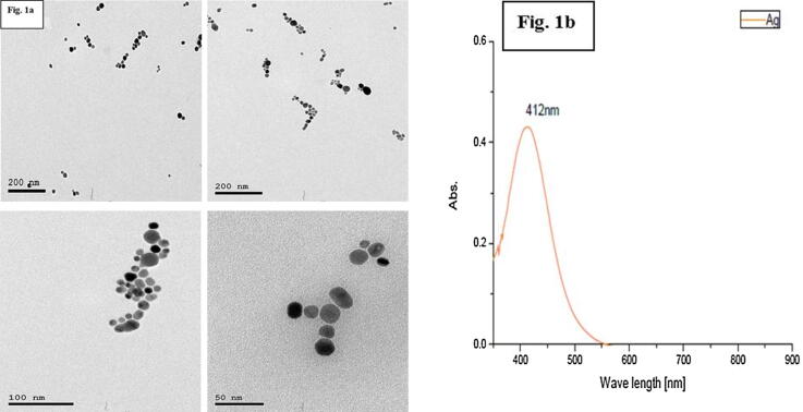
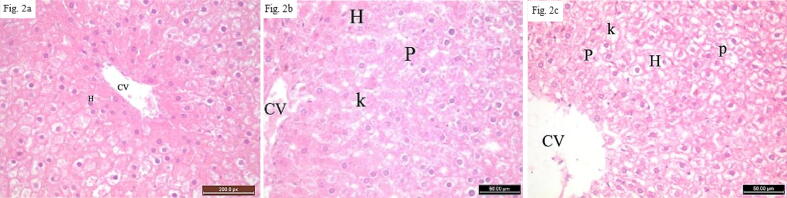
Products containing Silver nanoparticles (Ag NPs) are becoming vastly used in our daily life. The widespread increased introduction of Ag NPs in many aspects of life has raised researchers' concerns regarding their safety and toxicity for biological and environmental life in the past few years. The current study aimed to explore the subsequent effects of Ag NPs withdrawal, following short-term oral administration. Eighteen rats were assigned randomly into three groups (control group "1" and AG NPs treated groups "2" and "3"; 6 animals each). The control group received normal food and tap water while groups 2 & 3 received 0.5 ml of a solution containing 25 ppm Ag NPs for 14 days. Group 2 rats were sacrificed on day 14 whereas group 3 was left for another 14 days of particle cessation followed by euthanasia on day 28. Functional assessment was done by liver enzyme assays, hydrogen peroxide activity, hepatic Bdnf expression, and P53 immunoreactivity. Hepatic tissue structural assessment was done via hematoxylin and eosin, periodic acid-Schiff as well as Masson's trichrome stains. The results revealed a significant elevation of Hydrogen peroxide in group 2 only compared to the control group. Hepatic Bdnf and liver enzymes were both insignificantly affected. Structural abnormalities and enhanced apoptosis in hepatic tissue were found 14 days after ceasing the nanoparticles. In conclusion: Structural and functional insults following Ag NPs oral administration continues after particle withdrawal, and interestingly they do not necessitate apparent reflection on liver enzyme assays.
Keywords: ALT, Alanine Transaminase; AST, Aspartate Transaminase; Ag NPs, Silver Nanoparticles; Bdnf, Brain-derived neurotrophic factor; H&E, Hematoxylin & Eosin stain; H2o2, Hydrogen Peroxide; HCC, Hepatocellular Carcinoma; Hepatic Bdnf; Hepatotoxicity; Hydrogen peroxide; Liver enzymes; Masson's trichrome stains; NaBH4, Sodium Borohydride; Nanoparticles, (NPs); PAS, Periodic acid-Schiff; PVP, Polyvinyl Pyrrolidone; RNA, Ribonucleic acid; ROS, Reactive Oxygen Species; SGOT, Serum Glutamate oxaloacetate Transaminase; SGPT, Serum Glutamate pyruvate Transaminase; Silver Nanoparticles (Ag NPs); TEM, Transmission Electron Microscopy; TRI, Masson's trichrome stains; Transmission Electron Microscopy; UV–Vis, Ultraviolet–Visible Spectroscopy; Ultraviolet–Visible Spectroscopy; ppm, Parts Per Million.
© 2022 The Author(s).
Conflict of interest statement
The authors declare that they have no known competing financial interests or personal relationships that could have appeared to influence the work reported in this paper.
Figures


Similar articles
-
Impact of biosynthesized silver nanoparticles cytotoxicity on dental pulp of albino rats (histological and immunohistochemical study).J Oral Biol Craniofac Res. 2021 Jul-Sep;11(3):386-392. doi: 10.1016/j.jobcr.2021.04.002. Epub 2021 Apr 14. J Oral Biol Craniofac Res. 2021. PMID: 33996434 Free PMC article.
-
Therapeutic potential of silver nanoparticles from Helianthemum lippii extract for mitigating cadmium-induced hepatotoxicity: liver function parameters, oxidative stress, and histopathology in wistar rats.Front Bioeng Biotechnol. 2024 Jun 27;12:1400542. doi: 10.3389/fbioe.2024.1400542. eCollection 2024. Front Bioeng Biotechnol. 2024. PMID: 39007052 Free PMC article.
-
Modulation of liver and kidney toxicity by herb Withania somnifera for silver nanoparticles: a novel approach for harmonizing between safety and use of nanoparticles.Protoplasma. 2015 Mar;252(2):547-58. doi: 10.1007/s00709-014-0701-5. Epub 2014 Sep 24. Protoplasma. 2015. PMID: 25248758
-
Toxic Effects of Silver Nanoparticles on Liver and Some Hematological Parameters in Male and Female Mice (Mus musculus).Biol Trace Elem Res. 2015 Jun;165(2):153-8. doi: 10.1007/s12011-015-0247-1. Epub 2015 Feb 1. Biol Trace Elem Res. 2015. PMID: 25637567
-
A systematic review on silver nanoparticles-induced cytotoxicity: Physicochemical properties and perspectives.J Adv Res. 2017 Nov 2;9:1-16. doi: 10.1016/j.jare.2017.10.008. eCollection 2018 Jan. J Adv Res. 2017. PMID: 30046482 Free PMC article. Review.
Cited by
-
Do We Know Enough About the Safety Profile of Silver Nanoparticles in Oncology? A Focus on Novel Methods and Approaches.Int J Mol Sci. 2025 Jun 2;26(11):5344. doi: 10.3390/ijms26115344. Int J Mol Sci. 2025. PMID: 40508153 Free PMC article. Review.
References
-
- Aebi, H., 1984. [13] Catalase in vitro, in: Methods in Enzymology, Oxygen Radicals in Biological Systems. Academic Press, pp. 121–126. https://doi.org/10.1016/S0076-6879(84)05016-3
-
- Akter M., Sikder MdT., Rahman MdM., Ullah A.K.M.A., Hossain K.F.B., Banik S., Hosokawa T., Saito T., Kurasaki M. A systematic review on silver nanoparticles-induced cytotoxicity: physicochemical properties and perspectives. J. Adv. Res. 2018;9:1–16. doi: 10.1016/j.jare.2017.10.008. - DOI - PMC - PubMed
LinkOut - more resources
Full Text Sources
Research Materials
Miscellaneous
