. 2022 May;29(5):3568-3576.
doi: 10.1016/j.sjbs.2022.02.039. Epub 2022 Feb 26.

Hypoxia, a dynamic tool to amplify the gingival mesenchymal stem cells potential for neurotrophic factor secretion

Affiliations

Hypoxia, a dynamic tool to amplify the gingival mesenchymal stem cells potential for neurotrophic factor secretion

Shankargouda Patil et al. Saudi J Biol Sci. 2022 May.

Abstract

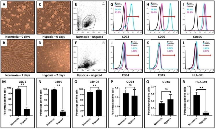
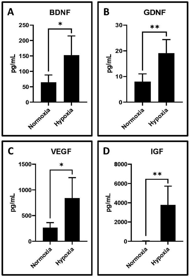
Gingival mesenchymal stem cells (GMSCs) have significant regenerative potential. Their potential applications range from the treatment of inflammatory diseases, wound healing, and oral disorders. Preconditioning these stem cells can optimize their biological properties. Hypoxia preconditioning of MSCs improves stem cell properties like proliferation, survival, and differentiation potential. This research explored the possible impact of hypoxia on the pluripotent stem cell properties that GMSCs possess. We evaluated the morphology, stemness, neurotrophic factors, and stemness-related genes. We compared the protein levels of secreted neurotrophic factors between normoxic and hypoxic GMSC-conditioned media (GMSC-CM). Results revealed that hypoxic cultured GMSC's had augmented expression of neurotrophic factors BDNF, GDNF, VEGF, and IGF1 and stemness-related gene NANOG. Hypoxic GMSCs showed decreased expression of the OCT4 gene. In hypoxic GMSC-CM, the neurotrophic factors secretions were significantly higher than normoxic GMSC-CM. Our data demonstrate that culturing of GMSCs in hypoxia enhances the secretion of neurotrophic factors that can lead to neuronal lineage differentiation.

Keywords: Gingival mesenchymal stem cell; Hypoxia; Neurotrophic factors; Normoxia.


Conflict of interest statement

The authors declare that they have no known competing financial interests or personal relationships that could have appeared to influence the work reported in this paper.

Figures

Fig. 1
Fig. 1
Isolation and characterization of GMSCs. (A) Photomicrograph of passage 2 with GMSCs. Scale bar = 100 μm. (B-H) Characterization of GMSCs for MSC-specific positive markers CD73, CD90, CD105 and negative markers CD34, CD45, HLA-DR. (GMSCs: Gingival mesenchymal stem cells).
Fig. 2
Fig. 2
Comparative characterization of GMSCs incubated at normoxia (∼20% oxygen) and hypoxia (0.5% – 1% oxygen) for MSC-specific markers. (A,B) Photomicrograph of GMSCs after normoxic incubation at day 0 and day 7. (C,D) Photomicrograph of GMSCs after hypoxic incubation at day 0 and day 7. Scale bar = 100 μm. (E-R) Comparative characterization of GMSCs incubated at normoxia and hypoxia for MSC-specific positive markers CD73, CD90, CD105 and negative markers CD34, CD45, HLA-DR. (ns: insignificant, **p < .01).
Fig. 3
Fig. 3
RT-qPCR analysis for gene expression in GMSCs incubated at normoxia and hypoxia. (A) Comparative gene expression (fold change) for gene expressing hypoxia-inducible factor (HIF-1α). (B-E) Comparative gene expression (fold change) for genes expressing neurotrophic factors BDNF, GDNF, VEGF, and IGF. (ns: not significant, **p < .01).
Fig. 4
Fig. 4
Western blot analysis for protein expression in GMSCs incubated at normoxia and hypoxia. (A, B) Comparative protein expression (optical density) for hypoxia-inducible factor (HIF-1α). (A, C-F) Comparative protein expression (optical density) for neurotrophic factors BDNF, GDNF, VEGF, and IGF. (**p < .01).
Fig. 5
Fig. 5
Bead-based flow cytometry analysis of secretome in GMSC-conditioned medium for secreted proteins by GMSCs incubated at normoxia and hypoxia. Comparative protein levels (pg/mL) for neurotrophic factors (A) BDNF, (B) GDNF, (C) VEGF, and (D) IGF. (*p < .05, **p < .01).
Fig. 6
Fig. 6
RT-qPCR analysis for gene expression in GMSCs incubated at normoxia and hypoxia. (A, B) Comparative gene expression (fold change) for genes expressing stemness-related transcription factors NANOG and OCT4. (*p < .05, **p < .01).

References

    1. Adesida A.B., Mulet-Sierra A., Jomha N.M. Hypoxia mediated isolation and expansion enhances the chondrogenic capacity of bone marrow mesenchymal stromal cells. Stem Cell Res. Ther. 2012;3:9. doi: 10.1186/scrt100. - DOI - PMC - PubMed
    1. Ahmed N.-E.-M.-B., Murakami M., Kaneko S., Nakashima M. The effects of hypoxia on the stemness properties of human dental pulp stem cells (DPSCs) Sci. Rep. 2016;6:35476. doi: 10.1038/srep35476. - DOI - PMC - PubMed
    1. Angelopoulos I., Brizuela C., Khoury M. Gingival Mesenchymal Stem Cells Outperform Haploidentical Dental Pulp-derived Mesenchymal Stem Cells in Proliferation Rate, Migration Ability, and Angiogenic Potential. Cell Transplant. 2018;27(6):967–978. doi: 10.1177/0963689718759649. - DOI - PMC - PubMed
    1. Antebi B., Rodriguez L.A., Walker K.P., Asher A.M., Kamucheka R.M., Alvarado L., Mohammadipoor A., Cancio L.C. Short-term physiological hypoxia potentiates the therapeutic function of mesenchymal stem cells. Stem Cell Res. Ther. 2018;9(1) doi: 10.1186/s13287-018-1007-x. - DOI - PMC - PubMed
    1. Aranha A.M.F., Zhang Z., Neiva K.G., Costa C.A.S., Hebling J., Nör J.E. Hypoxia Enhances the Angiogenic Potential of Human Dental Pulp Cells. J. Endod. 2010;36(10):1633–1637. doi: 10.1016/j.joen.2010.05.013. - DOI - PubMed