Reduced Immunity Regulator MAVS Contributes to Non-Hypertrophic Cardiac Dysfunction by Disturbing Energy Metabolism and Mitochondrial Homeostasis
- PMID: 35844503
- PMCID: PMC9283757
- DOI: 10.3389/fimmu.2022.919038
Reduced Immunity Regulator MAVS Contributes to Non-Hypertrophic Cardiac Dysfunction by Disturbing Energy Metabolism and Mitochondrial Homeostasis
Abstract
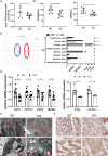
Cardiac dysfunction is manifested as decline of cardiac systolic function, and multiple cardiovascular diseases (CVDs) can develop cardiac insufficiency. Mitochondrial antiviral signaling (MAVS) is known as an innate immune regulator involved in viral infectious diseases and autoimmune diseases, whereas its role in the heart remains obscure. The alteration of MAVS was analyzed in animal models with non-hypertrophic and hypertrophic cardiac dysfunction. Then, MAVS-deficient mice were generated to examine the heart function, mitochondrial status and energy metabolism. In vitro, CRISPR/Cas9-based gene editing was used to delete MAVS in H9C2 cell lines and the phenotypes of mitochondria and energy metabolism were evaluated. Here we observed reduced MAVS expression in cardiac tissue from several non-hypertrophic cardiac dysfunction models, contrasting to the enhanced MAVS in hypertrophic heart. Furthermore, we examined the heart function in mice with partial or total MAVS deficiency and found spontaneously developed cardiac pump dysfunction and cardiac dilation as assessed by echocardiography parameters. Metabonomic results suggested MAVS deletion probably promoted cardiac dysfunction by disturbing energy metabolism, especially lipid metabolism. Disordered and mitochondrial homeostasis induced by mitochondrial oxidative stress and mitophagy impairment also advanced the progression of cardiac dysfunction of mice without MAVS. Knockout of MAVS using CRISPR/Cas9 in cardiomyocytes damaged mitochondrial structure and function, as well as increased mitochondrial ROS production. Therefore, reduced MAVS contributed to the pathogenesis of non-hypertrophic cardiac dysfunction, which reveals a link between a key regulator of immunity (MAVS) and heart function.
Keywords: MAVS; cardiac dysfunction; energy metabolism; mitochondrial dysfunction; oxidative stress.
Copyright © 2022 Wang, Sun, Cao, Lin, Wu, Li, Yin, Zhou, Huang, Zhang, Zhang, Xia and Jia.
Conflict of interest statement
The authors declare that the research was conducted in the absence of any commercial or financial relationships that could be construed as a potential conflict of interest.
Figures


Similar articles
-
Innate Immune Nod1/RIP2 Signaling Is Essential for Cardiac Hypertrophy but Requires Mitochondrial Antiviral Signaling Protein for Signal Transductions and Energy Balance.Circulation. 2020 Dec 8;142(23):2240-2258. doi: 10.1161/CIRCULATIONAHA.119.041213. Epub 2020 Oct 19. Circulation. 2020. PMID: 33070627
-
A conventional immune regulator mitochondrial antiviral signaling protein blocks hepatic steatosis by maintaining mitochondrial homeostasis.Hepatology. 2022 Feb;75(2):403-418. doi: 10.1002/hep.32126. Epub 2021 Dec 12. Hepatology. 2022. PMID: 34435375
-
Inducible Cardiac-Specific Deletion of Sirt1 in Male Mice Reveals Progressive Cardiac Dysfunction and Sensitization of the Heart to Pressure Overload.Int J Mol Sci. 2019 Oct 10;20(20):5005. doi: 10.3390/ijms20205005. Int J Mol Sci. 2019. PMID: 31658614 Free PMC article.
-
Effects of anticancer drugs on the cardiac mitochondrial toxicity and their underlying mechanisms for novel cardiac protective strategies.Life Sci. 2021 Jul 15;277:119607. doi: 10.1016/j.lfs.2021.119607. Epub 2021 May 13. Life Sci. 2021. PMID: 33992675 Review.
-
Mechanisms of MAVS regulation at the mitochondrial membrane.J Mol Biol. 2013 Dec 13;425(24):5009-19. doi: 10.1016/j.jmb.2013.10.007. Epub 2013 Oct 9. J Mol Biol. 2013. PMID: 24120683 Free PMC article. Review.
Cited by
-
Mitochondrial antiviral signaling protein: a potential therapeutic target in renal disease.Front Immunol. 2023 Oct 12;14:1266461. doi: 10.3389/fimmu.2023.1266461. eCollection 2023. Front Immunol. 2023. PMID: 37901251 Free PMC article. Review.
-
Metabolic profiling of urinary exosomes for systemic lupus erythematosus discrimination based on HPL-SEC/MALDI-TOF MS.Anal Bioanal Chem. 2023 Nov;415(26):6411-6420. doi: 10.1007/s00216-023-04916-z. Epub 2023 Aug 30. Anal Bioanal Chem. 2023. PMID: 37644324
-
Connection Between HIV and Mitochondria in Cardiovascular Disease and Implications for Treatments.Circ Res. 2024 May 24;134(11):1581-1606. doi: 10.1161/CIRCRESAHA.124.324296. Epub 2024 May 23. Circ Res. 2024. PMID: 38781302 Free PMC article. Review.
-
DDX3X/MAVS alleviates doxorubicin‑induced cardiotoxicity by regulating stress granules.Mol Med Rep. 2025 Sep;32(3):237. doi: 10.3892/mmr.2025.13602. Epub 2025 Jun 27. Mol Med Rep. 2025. PMID: 40576139 Free PMC article.
-
From mitochondria to heart: the role and challenges of mitochondrial antiviral signaling protein in cardiovascular disease.Front Cardiovasc Med. 2025 Aug 6;12:1572559. doi: 10.3389/fcvm.2025.1572559. eCollection 2025. Front Cardiovasc Med. 2025. PMID: 40842475 Free PMC article. Review.
References
-
- Caforio AL, Pankuweit S, Arbustini E, Basso C, Gimeno-Blanes J, Felix SB, et al. . Current State of Knowledge on Aetiology, Diagnosis, Management, and Therapy of Myocarditis: A Position Statement of the European Society of Cardiology Working Group on Myocardial and Pericardial Diseases. Eur Heart J (2013) 34(33):2636–48, 2648a-2648d. doi: 10.1093/eurheartj/eht210 - DOI - PubMed
Publication types
MeSH terms
Substances
LinkOut - more resources
Full Text Sources
Medical
Molecular Biology Databases
Miscellaneous

