Review
. 2022 Jun 30:13:926516.
doi: 10.3389/fimmu.2022.926516. eCollection 2022.

Key Factors for Thymic Function and Development

Affiliations
Review

Key Factors for Thymic Function and Development

Valentin P Shichkin et al. Front Immunol. .

Abstract

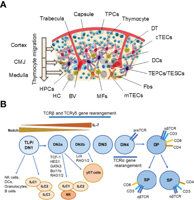
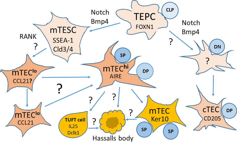
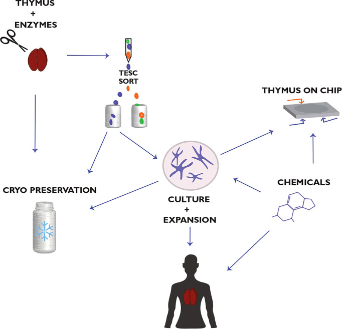
The thymus is the organ responsible for T cell development and the formation of the adaptive immunity function. Its multicellular environment consists mainly of the different stromal cells and maturing T lymphocytes. Thymus-specific progenitors of epithelial, mesenchymal, and lymphoid cells with stem cell properties represent only minor populations. The thymic stromal structure predominantly determines the function of the thymus. The stromal components, mostly epithelial and mesenchymal cells, form this specialized area. They support the consistent developmental program of functionally distinct conventional T cell subpopulations. These include the MHC restricted single positive CD4+ CD8- and CD4- CD8+ cells, regulatory T lymphocytes (Foxp3+), innate natural killer T cells (iNKT), and γδT cells. Several physiological causes comprising stress and aging and medical treatments such as thymectomy and chemo/radiotherapy can harm the thymus function. The present review summarizes our knowledge of the development and function of the thymus with a focus on thymic epithelial cells as well as other stromal components and the signaling and transcriptional pathways underlying the thymic cell interaction. These critical thymus components are significant for T cell differentiation and restoring the thymic function after damage to reach the therapeutic benefits.

Keywords: T cells; intrathymic regulators; thymic epithelial cells (TEC); thymic microenvironment; thymic stem cells; thymus; thymus regeneration.


Conflict of interest statement

Author VS was employed by company OmniFarma. The remaining author declares that the research was conducted in the absence of any commercial or financial relationships that could be construed as a potential conflict of interest.

Figures

Graphical Abstract
Graphical Abstract
Figure 1
Figure 1
Thymus cell architecture (A), and T cell and innate lymphoid cell (ILC) development in the thymus (B). The thymus consists of two lobes that are separated by connective tissue strands (trabeculae) in lobules. Each thymic lobule consisted of the cortex and medulla. The cortex contains CD34+ uncommitted pluripotent hematopoietic precursor cells (HPCs) entering the thymus at the cortico-medullary junction (CMJ) and migrating to the capsule, committed double negative (DN) CD4CD8 T precursor cells (TPCs) located in the subcapsular region (DN1–DN4 stages), and immature double positive (DP) CD4+CD8+ (Pre-DP) cortical thymocytes migrating through the cortex and CMJ to the medullar zone. The medulla contains single positive (SP) CD4+ and CD8+ naïve thymocytes migrating to the periphery after maturing. Stromal-epithelial compartment of the thymus is submitted by minor populations of EpCam+ (CD326+) Foxn1+ bipotent thymic epithelial precursor cells/thymic epithelial stem cells (TEPCs/TESCs), and mesenchymal stem cells (MSCs) located probably into the thymic parenchyma close to the CMJ region, as well as EpCam+CD205+ cortical thymic epithelial cells (cTECs) located in the cortex and EpCam+Air+ medullary thymic epithelial cells (mTECs) located in the medulla. The cortex and medulla also contain macrophages (MFs), fibroblasts (Fbs), and dendritic cells (DCs) that, together with cTECs and mTECs, participate in the differentiation, maturation, and positive and negative selection of thymocytes. T cell and ILC lineages diverge at the stages of early T precursors/double negative 1 (ETP/DN1) and the DN2-DN3 transition stage. Depending on the status of the TCR loci, the strength of Notch signaling and activities of E-ld proteins and Bcl11b, multipotent TLPs may develop conventional αβ T cells or acquire innate-like properties and give rise to thymic natural killer (NK) cells, DCs, granulocytes, B cells, one of three ILC subsets and invariant γδ T cells. Resident ILC progenitors have been suggested to originate from failed T cell development and locally maintain the mature ILC pool. BV, Blood Vessel; DT, Dead Thymocytes; HC, Hassall’s Corpuscle. (A) modified from Shichkin and Antica, 2020 (9); the article is licensed under a Creative Commons Attribution 4.0 International License. (B) modified from Shin and McNagny, 2021 (138); the article is distributed under the terms of the Creative Commons Attribution License (CC BY).
Figure 2
Figure 2
Key markers and pathways in the development of thymic epithelial cells (TECs) from bipotent thymic epithelial progenitor cells (TEPCs). TEPCs differentiate into medullary and cortical thymic epithelial cell lineages (mTECs and cTECs, respectively) that are regulated by Foxn1 expression. mTEC development goes through an intermediate stem cell stage (mTESCs), expressing the stem cell marker SSEA-1, and requires Notch signaling for the formation of mature Aire expressing mTECshi. Other pathways to the differentiation of mTEC subsets are still a matter of intensive research. It is also yet not known whether cTEC development goes through a similar intermediate stage. CLP, Committed Lymphocyte Precursor; DN, Double Negative Thymocytes; DP, Double Positive Thymocytes; SP, Single Positive Thymocytes. Modified from Alawam et al., 2020 (71); the article is distributed under the terms of the Creative Commons Attribution License (CC BY).
Figure 3
Figure 3
Thymic epithelial stem cell (TESC)-based strategy for thymus regeneration with small chemical compounds (SCC). This strategy proposes the collection, preparation, and cryopreservation of primary thymic tissue and TESC-enriched cell samples. These thymic samples are used further to select TESC-specific SCC that can regulate the differentiation and proliferation of human TESCs and support their clonal expansion. The selected SCC are tested also for supporting thymic tissue growth in vitro as well as for reconstitution of thymic function in terms of differentiation, maturation, and tolerance of autologous T cells. The use of microfluidic chips in combination with human 3D thymic organ cultures (Thymus-on-Chip devices) to assess SCC specificity and toxicity is essential to accelerate drug development for thymus-compromised patients. Actual challenges are optimizing the thymectomy procedure in patients to preserve a thymic fragment for consequent postsurgical thymus regeneration and the quality life monitoring of thymectomized patients concerning their resistance to infections, allergies, autoimmune, oncological, and other diseases associated with the impaired thymic function. Modified from Shichkin and Antica, 2020 (9); the article is licensed under a Creative Commons Attribution 4.0 International License.

Similar articles

Cited by

References

    1. Gordon J, Manley NR. Mechanisms of Thymus Organogenesis and Morphogenesis. Development (2011) 138:386578. doi: 10.1242/dev.059998 - DOI - PMC - PubMed
    1. Muсoz JJ, Zapata AG. “Thymus Ontogeny and Development”. In: Passos GA, editor. Thymus Transcriptome and Cell Biology. Springer Nature Switzerland AG, Cham: Springer Nature Switzerland AG; (2019). p. 19–34. doi: 10.1007/978-3-030-12040-5_2 - DOI
    1. Onder L, Nindl V, Scandella E, Chai Q, Cheng HW, Caviezel-Firner S, et al. Alternative NF-Kappa B Signaling Regulates mTEC Differentiation From Podoplanin-Expressing Presursors in the Cortico-Medullary Junction. Eur J Immunol (2015) 45:2218–31. doi: 10.1002/eji.201545677 - DOI - PubMed
    1. Nitta T, Takayanagi H. Non-Epithelial Thymic Stromal Cells: Unsung Heroes in Thymus Organogenesis and T Cell Development. Front Immunol (2021) 11:620894. doi: 10.3389/fimmu.2020.620894 - DOI - PMC - PubMed
    1. Skogberg G, Lundberg V, Berglund M, Gudmundsdottir J, Telemo E, Lindgren S, et al. Human Thymic Epithelial Primary Cells Produce Exosomes Carrying Tissue-Restricted Antigens. Immunol Cell Biol (2015) 93:72734. doi: 10.1038/icb.2015.33 - DOI - PMC - PubMed

Publication types