. 2022 Jun 30:13:884760.
doi: 10.3389/fimmu.2022.884760. eCollection 2022.

Immunogenicity of SARS-CoV-2 Trimeric Spike Protein Associated to Poly(I:C) Plus Alum

Affiliations

Immunogenicity of SARS-CoV-2 Trimeric Spike Protein Associated to Poly(I:C) Plus Alum

Júlio Souza Dos-Santos et al. Front Immunol. .

Abstract

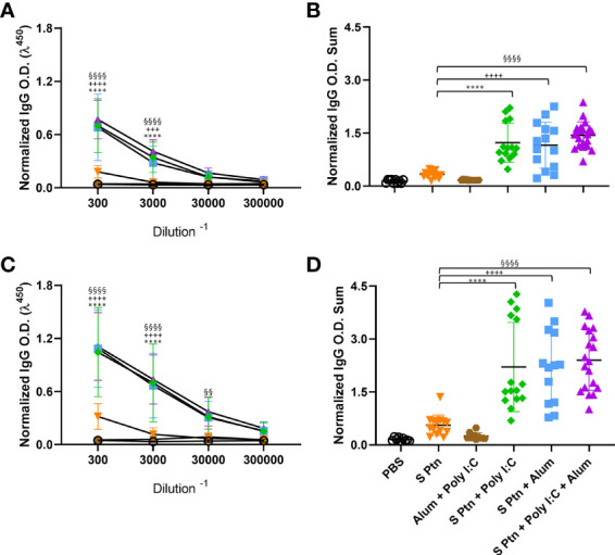
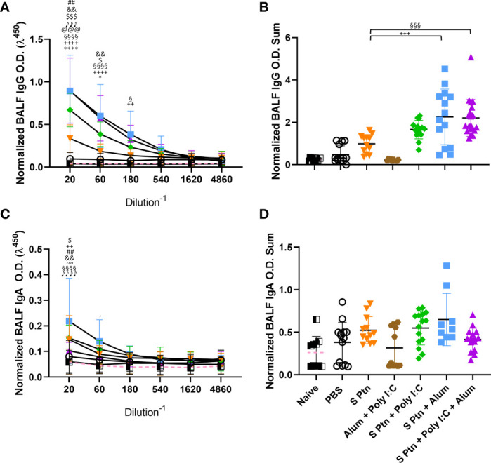
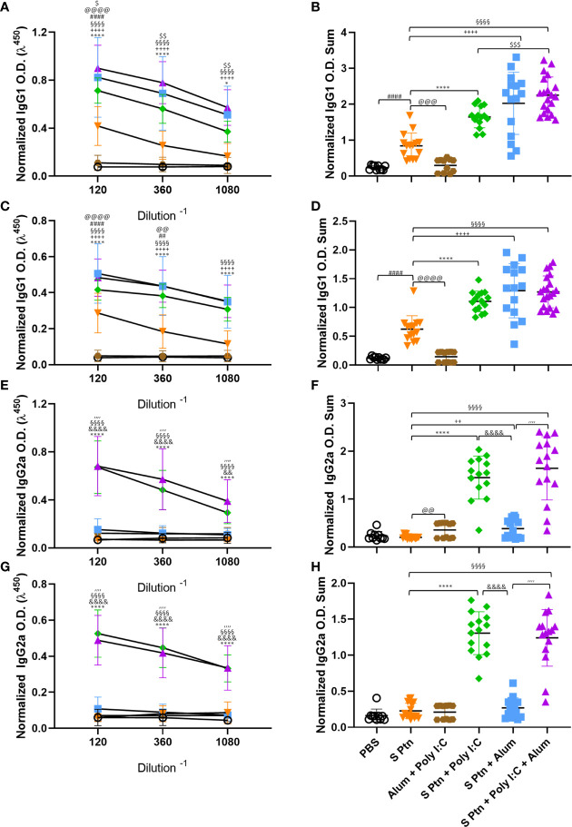
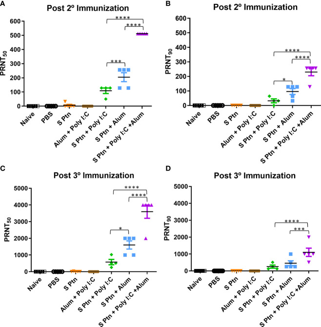
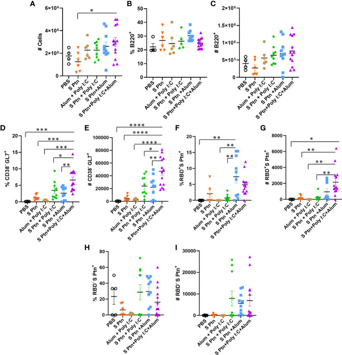
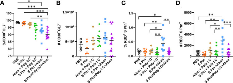
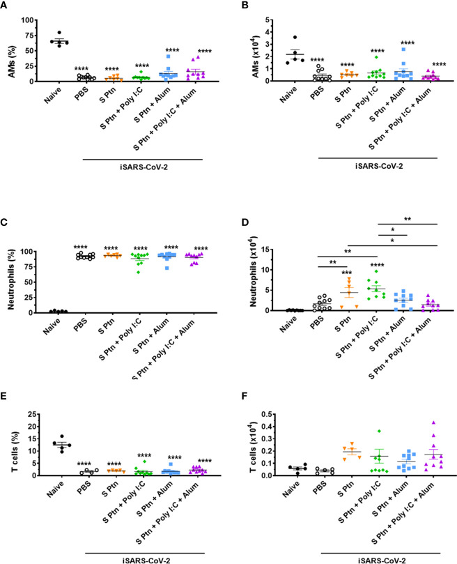
The SARS-CoV-2 pandemic has had a social and economic impact worldwide, and vaccination is an efficient strategy for diminishing those damages. New adjuvant formulations are required for the high vaccine demands, especially adjuvant formulations that induce a Th1 phenotype. Herein we assess a vaccination strategy using a combination of Alum and polyinosinic:polycytidylic acid [Poly(I:C)] adjuvants plus the SARS-CoV-2 spike protein in a prefusion trimeric conformation by an intradermal (ID) route. We found high levels of IgG anti-spike antibodies in the serum by enzyme linked immunosorbent assay (ELISA) and high neutralizing titers against SARS-CoV-2 in vitro by neutralization assay, after two or three immunizations. By evaluating the production of IgG subtypes, as expected, we found that formulations containing Poly(I:C) induced IgG2a whereas Alum did not. The combination of these two adjuvants induced high levels of both IgG1 and IgG2a. In addition, cellular immune responses of CD4+ and CD8+ T cells producing interferon-gamma were equivalent, demonstrating that the Alum + Poly(I:C) combination supported a Th1 profile. Based on the high neutralizing titers, we evaluated B cells in the germinal centers, which are specific for receptor-binding domain (RBD) and spike, and observed that more positive B cells were induced upon the Alum + Poly(I:C) combination. Moreover, these B cells produced antibodies against both RBD and non-RBD sites. We also studied the impact of this vaccination preparation [spike protein with Alum + Poly(I:C)] in the lungs of mice challenged with inactivated SARS-CoV-2 virus. We found a production of IgG, but not IgA, and a reduction in neutrophil recruitment in the bronchoalveolar lavage fluid (BALF) of mice, suggesting that our immunization scheme reduced lung inflammation. Altogether, our data suggest that Alum and Poly(I:C) together is a possible adjuvant combination for vaccines against SARS-CoV-2 by the intradermal route.

Keywords: SARS-CoV-2; adjuvants; alum; intradermal route; poly (I:C); spike protein; vaccine.


Conflict of interest statement

The authors declare that the research was conducted in the absence of any commercial or financial relationships that could be construed as a potential conflict of interest.

Figures

Figure 1
Figure 1
Formulations containing adjuvants induce strong IgG response in the serum. Antigen-specific antibody levels were determined by ELISA and normalized using the control groups. Serum IgG levels were evaluated after two (A, B) or three immunizations (C, D). The summatory of all dilutions are represented in (B, D). Data in this figure consists 4 independent experiments normalized using the control groups and shown as mean ± S.D. Groups: Naïve (n=10); PBS (n=15); S Ptn (n=15); Alum + Poly(I:C) (n=11); S Ptn + Poly(I:C) (n=15); S Ptn + Alum (n=15); S Ptn + Poly(I:C) + Alum (n=20). * - represents differences between S Ptn and S Ptn + Poly(I:C) groups; + - represents differences between S Ptn and S Ptn + Alum groups; § - represents differences between S Ptn and S Ptn + Poly(I:C) + Alum groups. (A, C) was performed two-way ANOVA followed by Bonferroni post-test and (B, D) was analyzed by one-way ANOVA with Tukey’s post hoc test. ****p<0.0001.
Figure 2
Figure 2
Formulations containing adjuvants induce a strong IgG, but not IgA, response in the BALF. Antigen-specific antibody levels in the BALF were determined by ELISA and normalized using the control groups. Twenty-four hours after the viral challenge we euthanized mice and evaluated in the BALF the IgG (A, B) and IgA levels (C, D) were determined. The summatory of all dilutions are represented in (B, D). Data in this figure consists 4 independent experiments normalized using the control groups and shown as mean ± S.D. Groups: Naïve (n=10); PBS (n=15); S Ptn (n=15); Alum + Poly(I:C) (n=11); S Ptn + Poly(I:C) (n=15); S Ptn + Alum (n=15); S Ptn + Poly(I:C) + Alum (n=20). # - represents differences between PBS and S Ptn groups; @ - represents differences between S Ptn and Alum + Poly(I:C) groups; * - represents differences between S Ptn and S Ptn + Poly(I:C) groups; + - represents differences between S Ptn and S Ptn + Alum groups; § - represents differences between S Ptn and S Ptn + Poly(I:C) + Alum groups; $ - represents differences between S Ptn + Poly(I:C) and S Ptn + Poly(I:C) + Alum groups; & - represents differences between S Ptn + Poly(I:C) and S Ptn + Alum groups; ‘ - represents differences between S Ptn + Alum and S Ptn + Poly(I:C) + Alum groups; ♪ - represents differences between Naïve and S Ptn groups. (A, C) was performed two-way ANOVA followed by Bonferroni post-test and (B, D) was analyzed by one-way ANOVA with Tukey’s post hoc test. *p<0.05, ****p<0.0001.
Figure 3
Figure 3
Formulations containing Poly(I:C) were able to induce type 1 response serum antibodies. Antigen-specific antibody levels were determined by ELISA and normalized using the control groups. Serum IgG1 (A–D) and IgG2a (E–H) levels were evaluated after two (A, B, E, F) or three immunizations (C, D, G, H). The summatory of all dilutions are represented in (B, D, F, H). Data in this figure consists 4 independent experiments normalized using the control groups and shown as mean ± S.D. Groups: PBS (n=10); S Ptn (n=15); Alum + Poly(I:C) (n=11); S Ptn + Poly(I:C) (n=15); S Ptn + Alum (n=15); S Ptn + Poly(I:C) + Alum (n=20). # - represents differences between PBS and S Ptn groups; @ - represents differences between S Ptn and Alum + Poly(I:C) groups; * - represents differences between S Ptn and S Ptn + Poly(I:C) groups; + - represents differences between S Ptn and S Ptn + Alum groups; § - represents differences between S Ptn and S Ptn + Poly(I:C) + Alum groups; $ - represents differences between S Ptn + Poly(I:C) and S Ptn + Poly(I:C) + Alum groups; & - represents differences between S Ptn + Poly(I:C) and S Ptn + Alum groups; ‘ - represents differences between S Ptn + Alum and S Ptn + Poly(I:C) + Alum groups. (A, C, E, G) was performed two-way ANOVA followed by Bonferroni post-test and (B, D, F, H) was analyzed by one-way ANOVA with Tukey’s post hoc test. *p<0.05, ****p<0.0001.
Figure 4
Figure 4
Spike protein associated to Poly(I:C) plus Alum co-administrated induced high titers of neutralizing antibodies. Titers of neutralizing antibodies were determined in vitro by neutralization assay after two and three immunizations. PRNT50 (A, C) and PRNT90 (B, D) for mice plasma collected 7 days after two and three immunization. Figure representative of 4 independent experiments and shown as mean ± S.D. and analyzed by one-way ANOVA with Tukey’s post hoc test. *p<0.05, ***p<0.001, ****p<0.0001.
Figure 5
Figure 5
B cell response within the germinal center after immunization. Lymphocytes from the draining popliteal lymph node were analyzed after three immunizations intradermal immunization with S protein alone or with adjuvants [Poly(I:C); Alum; Poly(I:C) + Alum]. Controls were performed with PBS or Poly(I:C) + Alum. (A) Number of total cells. (B) Percentage of B220+ cells. (C) Number of B220+ cells. (D) Percentage of CD38+GL7- cells. (E) Number of CD38+GL7- cells. (F) Percentage of RBD+S Ptn+ cells. (G) Number of RBD+S Ptn+ cells. (H) Percentage of RBD-S Ptn+ cells. (I) Number of RBD-S Ptn+ cells. Figure representative of 4 independent experiments and shown as mean ± S.D. and was performed by one-way ANOVA with Tukey’s post hoc test. *p<0.03, **p<0.005, ***p<0.0002, ****p<0.0001. (SEM; n=4-13).
Figure 6
Figure 6
B cell profile outside the germinal center after immunization. After three immunizations, lymphocytes from the draining popliteal lymph node macerated after intradermal immunization with S protein alone or with adjuvants [Poly(I:C); Alum; Poly(I:C) + Alum]. Controls were performed with PBS or Poly(I:C) + Alum. (A) Percentage of CD38+GL7- cells. (B) Number of CD38+GL7- cells. (C) Percentage of RBD+S Ptn+ cells. (D) Number of RBD+S Ptn+ cells. Figure representative of 4 independent experiments and shown as mean ± S.D. and was performed by one-way ANOVA with Tukey’s post hoc test. *p<0.05, **p<0.005, ***p<0.0005. (SEM; n=4-13).
Figure 7
Figure 7
Analysis of BALF immune cell content in mice immunized with S Ptn together with different adjuvants following challenge with inactivated SARS-CoV-2 virus. BALF cells were collected 24 hours after inactivated SARS-CoV-2 (iSARS-CoV-2) virus challenge. The percentage and number of AMs, neutrophils, and T cells from naïve and mice immunized with S Ptn and different adjuvant combinations as gated on flow cytometry plots using markers including SiglecF, CD11c, CD11b, Ly6G, Ly6C, and TCRβ. Percentage of (A) AMs (CD11b+CD11c-Ly6chiF4/80+), (C) Neutrophils (CD11b+CD11c-Ly6c+F4/80-), and (E) T cells (CD11b+CD11c-Ly6c+F4/80-) in the BALFs. Absolute numbers of (B) AMs, (D) Neutrophils, and (F) T cells in the BALFs. Data are presented as the mean ± SEM of three pooled experiments and analyzed by one-way ANOVA with Tukey’s post hoc test, *p<0.05, **p<0.01, ***p<0.001, ****p<0.0001.

Similar articles

Cited by

References

    1. Carter D, van Hoeven N, Baldwin S, Levin Y, Kochba E, Magill A, et al. . The Adjuvant GLA-AF Enhances Human Intradermal Vaccine Responses. Sci Adv (2018) 4(9):eaas9930. doi: 10.1126/sciadv.aas9930 - DOI - PMC - PubMed
    1. Tebas P, Kimberly AK, Ami P, Joel NM, Matthew PM, Albert JS, et al. . Intradermal Syncon® Ebola GP DNA Vaccine Is Temperature Stable And Safely Demonstrates Cellular And Humoral Immunogenicity Advantages In Healthy Volunteers. J Of Infect Dis (2019) 220(3):400–10. doi: 10.1093/infdis/jiz132 - DOI - PubMed
    1. Frey SE, Anna W, Srilatha E, Lisa AJ, Jack TS, Hana ES, et al. . Comparison Of Lyophilized Versus Liquid Modified Vaccinia Ankara (Mva) Formulations and Subcutaneous Versus Intradermal Routes Of Administration In Healthy Vaccinia-Naïve Subjects”. Vaccine (2015) 33(39):5225–34. doi: 10.1016/j.vaccine.2015.06.075 - DOI - PMC - PubMed
    1. Tebas P, ShuPing Y, Jean DB, Emma LR, Ami P, Aaron CQ, et al. . Safety and Immunogenicity of INO-4800 DNA Vaccine Against SARS-Cov-2: A Preliminary Report of an Open-Label, Phase 1 Clinical Trial. EClinicalMedicine (2021) 31:100689. doi: 10.1016/j.eclinm.2020.100689 - DOI - PMC - PubMed
    1. Zeng F, Chow KY, Hon CC, Law KM, Yip CW, Chan KH, et al. . Characterization of Humoral Responses in Mice Immunized With Plasmid DNAs Encoding SARS-CoV Spike Gene Fragments. Biochem Biophys Res Commun (2004) 315(4):1134–9. doi: 10.1016/j.bbrc.2004.01.166 - DOI - PMC - PubMed

Publication types