. 2022 Jun 29:13:913955.
doi: 10.3389/fimmu.2022.913955. eCollection 2022.

The Non-Receptor Protein Tyrosine Phosphatase PTPN6 Mediates a Positive Regulatory Approach From the Interferon Regulatory Factor to the JAK/STAT Pathway in Litopenaeus vannamei

Affiliations

The Non-Receptor Protein Tyrosine Phosphatase PTPN6 Mediates a Positive Regulatory Approach From the Interferon Regulatory Factor to the JAK/STAT Pathway in Litopenaeus vannamei

Mengting Luo et al. Front Immunol. .

Abstract

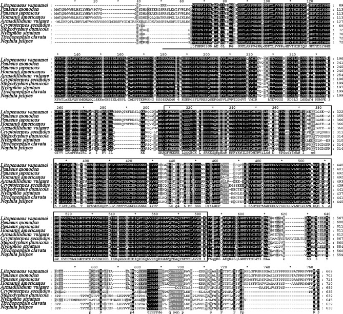
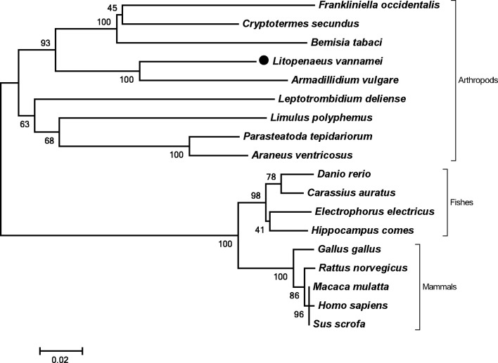
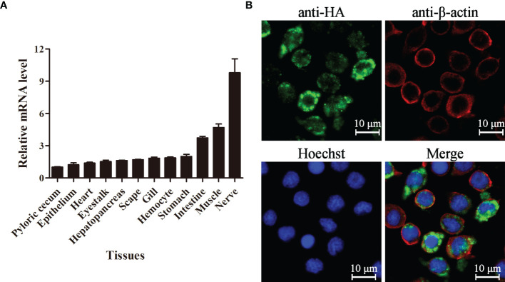
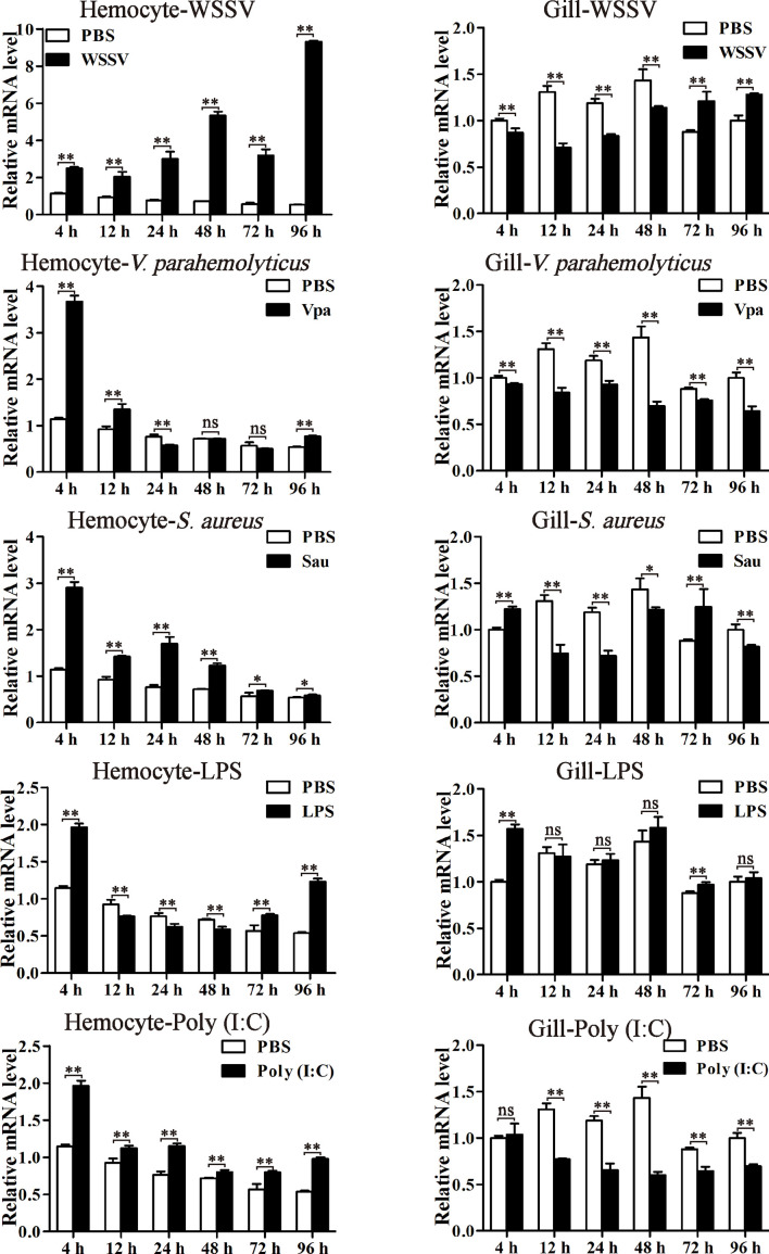
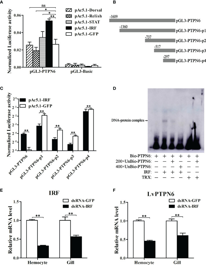
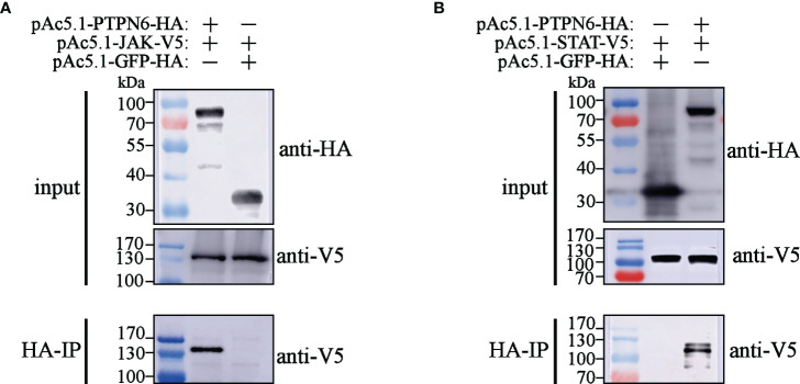
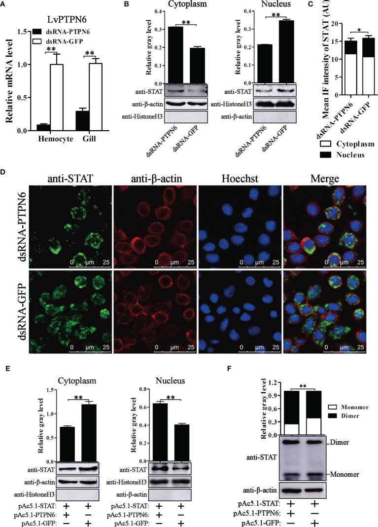
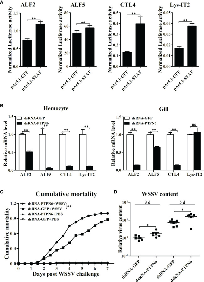
SH2-domain-containing protein tyrosine phosphatases (PTPs), belonging to the class I PTP superfamily, are responsible for the dephosphorylation on the phosphorylated tyrosine residues in some proteins that are involved in multiple biological processes in eukaryotes. The Janus kinase/signal transducers and activators of transcription (JAK/STAT) pathway transduce signaling responding to interferons and initiate cellular antiviral responses. The activity of the JAK/STAT pathway is generally orchestrated by the de-/phosphorylation of the tyrosine and serine residues of JAKs and STATs, in which the dephosphorylation processes are mainly controlled by PTPs. In the present study, an SH2-domian-contianing PTP, temporally named as LvPTPN6, was identified in Litopenaeus vannamei. LvPTPN6 shares high similarity with PTPN6s from other organisms and was phylogenetically categorized into the clade of arthropods that differs from those of fishes and mammals. LvPTPN6 was constitutively expressed in all detected tissues, located mainly in the cytoplasm, and differentially induced in hemocyte and gill after the challenge of stimulants, indicating its complicated regulatory roles in shrimp immune responses. Intriguingly, the expression of LvPTPN6 was regulated by interferon regulatory factor (IRF), which could directly bind to the LvPTPN6 promoter. Surprisingly, unlike other PTPN6s, LvPTPN6 could promote the dimerization of STAT and facilitate its nuclear localization, which further elevated the expression of STAT-targeting immune effector genes and enhanced the antiviral immunity of shrimp. Therefore, this study suggests a PTPN6-mediated regulatory approach from IRF to the JAK/STAT signaling pathway in shrimp, which provides new insights into the regulatory roles of PTPs in the JAK/STAT signaling pathway and contributes to the further understanding of the mechanisms of antiviral immunity in invertebrates.

Keywords: IFN regulatory factor; JAK/STAT signaling pathway; Litopenaeus vannamei; antiviral immunity; non-receptor protein tyrosine phosphatase.


Conflict of interest statement

The authors declare that the research was conducted in the absence of any commercial or financial relationships that could be construed as a potential conflict of interest.

Figures

Figure 1
Figure 1
Multiple-sequence alignment of PTPN6 homologs. The two predicted SH2 domains were underlined, and the PTPc domain was framed. The amino acid sequence of PTPN6s was obtained from NCBI with GenBank accession numbers of Penaeus monodon (XP_037785856.1), Penaeus japonicas (XP_042879519.1), Homarus americanus (XP_042242946.1), Armadillidium vulgare (RXG72814.1), Cryptotermes secundus (XP_023713602.1), Stegodyphus dumicola (XP_035233634.1), Nymphon striatum (KAG1683061.1), Trichonephila clavata (GFR09275.1), and Nephila pilipes (GFS80663.1). The five tines star "*" means the middle position of the two Numbers beside it.
Figure 2
Figure 2
Phylogenetic tree of PTPN6s. Amino acid sequences of PTPN6s were obtained from NCBI with GenBank accession numbers of Frankliniella occidentalis (XP_026279043.1), Cryptotermes secundus (XP_023713602.1), Bemisia tabaci (XP_018912318.1), Armadillidium vulgare (RXG72814.1), Leptotrombidium deliense (RWS28580.1), Limulus polyphemus (XP_022245683.1), Parasteatoda tepidariorum (XP_015930014.1), Araneus ventricosus (GBM95823.1), Danio rerio (NP_956140.1), Carassius auratus (XP_026121403.1), Electrophorus electricus (XP_026888060.1), Hippocampus comes (XP_019712005.1), Gallus gallus (NP_990299.1), Rattus norvegicus (NP_001171064.1), Macaca mulatta (NP_001248038.1), Homo sapiens (NP_002822), and Sus scrofa (XP_013845818.1).
Figure 3
Figure 3
Tissue distribution and subcellular localization of LvPTPN6. (A) Expression of LvPTPN6 in L. vannamei tissues detected by qRT-PCR with EF-1α as internal control. The expression level of LvPTPN6 in pyloric cecum was set as the baseline (1.0). Each bar represents the mean ± SD (n = 4). (B) Subcellular localization of HA-tagged LvPTPN6 was detected by confocal laser scanning microscopy analysis in S2 cells. LvPTPN6 was stained with Alexa Fluor 488 (green), the cytomembranes were visualized by β-actin stain with Alexa Flour 594 (red), and the nuclei were stained with Hoechst 33342 (blue).
Figure 4
Figure 4
The expression profiles of LvPTPN6 after immune stimulation. The expression of LvPTPN6 in hemocyte and gill of WSSV-, V. parahaemolyticus- (Vpa), S. aureus- (Sau), LPS-, and Poly(I:C)-challenged shrimps were detected using qRT-PCR. In each panel, the value of PBS group at 4 h was set as the baseline (1.0). Each bar represents the mean ± SD (n = 4), **p < 0.01, *p < 0.05, and ns > 0.05 by two-tailed unpaired Student’s t-test.
Figure 5
Figure 5
Regulation of LvPTPN6 expression by IRF. (A) Regulatory effects of Dorsal, Relish, STAT, IRF, and GFP (as control) on the LvPTPN6 promoter. Each bar represents the mean ± SD (n = 8), **p < 0.01, *p < 0.05, and ns: P > 0.05 by two-tailed unpaired Student’s t-test. (B) Scheme of the cleaved promoters of LvPTPN6. (C) Regulatory effects of IRF and GFP on the cleaved promoters of LvPTPN6. Each bar represents the mean ± SD (n = 8), **p < 0.01 by two-tailed unpaired Student’s t-test. (D) Interaction of IRF with the LvPTPN6 promoter analyzed by EMSA. The biotin-labeled (Bio-) or unlabeled (Unbio-) probes and purified prokaryotic protein of LvPTPN6 or TRX (as control) are used. (E, F) qRT-PCR analysis of IRF and LvPTPN6 mRNA levels in hemocyte and gill at 48 h post dsRNA injection. Values in the dsRNA-GFP control group were set as the baseline (1.0). Each bar represents the mean ± SD (n = 4), **p < 0.01 by two-tailed unpaired Student’s t-test.
Figure 6
Figure 6
Co-IP analysis of the interaction between LvPTPN6 and JAK/STAT. Interaction between HA-tagged LvPTPN6 and V5-tagged JAK (A) or STAT (B), HA-tagged GFP was set as internal control. Interacted proteins were precipitated by anti-HA affinity agarose.
Figure 7
Figure 7
Nuclear localization of STAT regulated by LvPTPN6. (A) qRT-PCR analysis of the LvPTPN6 mRNA level in hemocyte and gill at 48 h post dsRNA injection. Values in the dsRNA-GFP control group were set as the baseline (1.0). Each bar represents the mean ± SD (n = 4), **p < 0.01 by two-tailed unpaired Student’s t-test. (B) Western blot analysis of the protein level of STAT located in the cytoplasm and nucleus of hemocytes after dsRNA injection. (C) Immunofluorescence intensities (arbitrary units, AU) of cytoplasm- and nuclear-localized STAT in dsNRA-GFP- and dsRNA-PTPN6-treated hemocytes. Immunofluorescence intensities were calculated using JACoP with an ImageJ plugin from four randomly selected microscopic vision fields ( Supplemental Figure 3 ). *p < 0.05 by two-tailed unpaired Student’s t-test. (D) Immunofluorescent analysis of STAT in hemocytes after dsRNA injection. STAT was stained with Alexa Fluor 488 (green), the cytomembranes were visualized by β-actin stain with Alexa Flour 594 (red), and the nuclei were stained with Hoechst 33342 (blue). (E) Western blot analysis of the protein level of shrimp STAT located in the cytoplasm and nucleus of S2 cells overexpressed with LvPTPN6 or GFP (negative control). (B, E) In cytoplasm, the gray values of STAT bands were normalized to those of the cytoplasmic internal control of β-actin, and Histone H3 was detected to verify no contamination of nuclear protein. In nucleus, the gray values of STAT bans were normalized to those of the nuclear internal control of Histone H3, and β-actin was detected to verify no contamination of cytoplasmic protein. (F) Western blot analysis of the dimer and monomer levels of STAT through native PAGE after overexpressing LvPTPN6 in S2 cells. The gray values of STAT bans were normalized to those of the internal control of β-actin. (B–F) Each bar is mean ± SD of three independent quantification of the electrophoretic bands, **p < 0.01 by two-tailed unpaired Student’s t-test.
Figure 8
Figure 8
Role of LvPTPN6 in shrimp antiviral immunity. (A) Regulatory effects of shrimp STAT on the promoter of immune effector genes. Each bar represents the mean ± SD (n = 8), **p < 0.01 by two-tailed unpaired Student’s t-test. (B) qRT-PCR analysis of the mRNA level of STAT-regulated immune effector genes in hemocyte and gill at 48 h after dsRNA injection. Values in the dsRNA-GFP control group were set as the baseline (1.0). Each bar represents the mean ± SD (n = 4), **p < 0.01 and ns: P > 0.05 by two-tailed unpaired Student’s t-test. (C) Cumulative mortality of dsRNA-injected shrimps (n = 40) after WSSV infection. Data were recorded every 6 h and statistically analyzed by Kaplan–Meier log-rank χ2 test, **p < 0.01. (D) The relative viral load in muscle was analyzed by detecting the DNA of the WSSV ie1 gene in six randomly selected shrimps using qRT-PCR with three repetitions, and the shrimp EF-1α gene was used as the internal control. The level in dsRNA-GFP group at 3 days post WSSV infection was set as baseline (1.0). *p < 0.05 by two-tailed unpaired Student’s t-test.

Similar articles

Cited by

References

    1. Mustelin T, Feng GS, Bottini N, Alonso A, Kholod N, Birle D, et al. . Protein Tyrosine Phosphatases. Front Biosci (2002) 7:d85–142. doi: 10.2741/mustelin - DOI - PubMed
    1. Fischer EH, Charbonneau H, Tonks NK. Protein Tyrosine Phosphatases: A Diverse Family of Intracellular and Transmembrane Enzymes. Science (1991) 253:401–06. doi: 10.1126/science.1650499 - DOI - PubMed
    1. Alonso A, Sasin J, Bottini N, Friedberg I, Friedberg I, Osterman A, et al. . Protein Tyrosine Phosphatases in the Human Genome. Cell (2004) 117:699–711. doi: 10.1016/j.cell.2004.05.018 - DOI - PubMed
    1. Hasegawa K, Martin F, Huang G, Tumas D, Diehl L, Chan AC. Pest Domain-Enriched Tyrosine Phosphatase (PEP) Regulation of Effector/Memory T Cells. Science (2004) 303:685–89. doi: 10.1126/science.1092138 - DOI - PubMed
    1. Andersen JN, Jansen PG, Echwald SM, Mortensen OH, Fukada T, Del VR, et al. . A Genomic Perspective on Protein Tyrosine Phosphatases: Gene Structure, Pseudogenes, and Genetic Disease Linkage. FASEB J (2004) 18:8–30. doi: 10.1096/fj.02-1212rev - DOI - PubMed

Publication types

LinkOut - more resources