

Neutralizing Potency of Prototype and Omicron RBD mRNA Vaccines Against Omicron Variant
- PMID: 35844601
- PMCID: PMC9280631
- DOI: 10.3389/fimmu.2022.908478
Neutralizing Potency of Prototype and Omicron RBD mRNA Vaccines Against Omicron Variant
Abstract
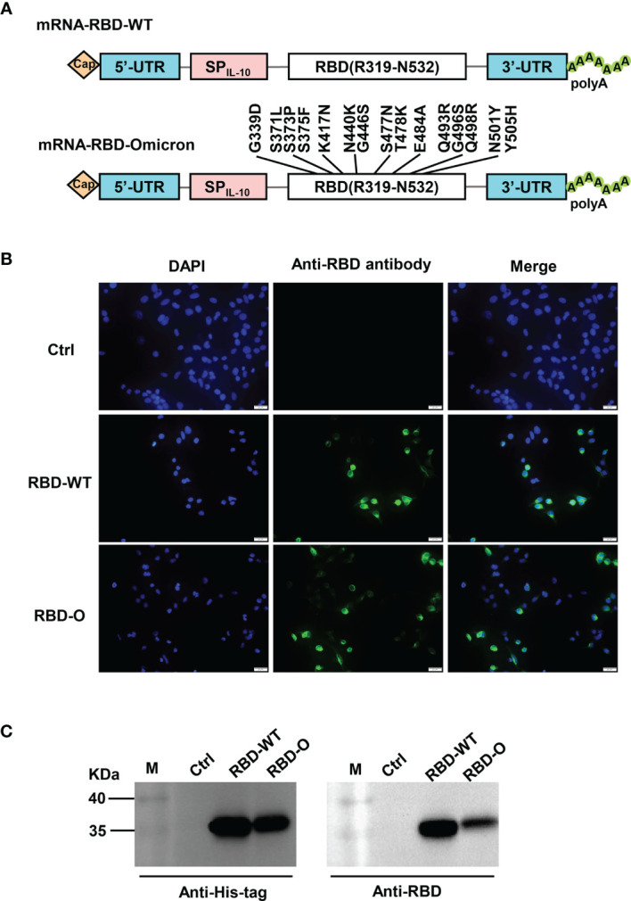
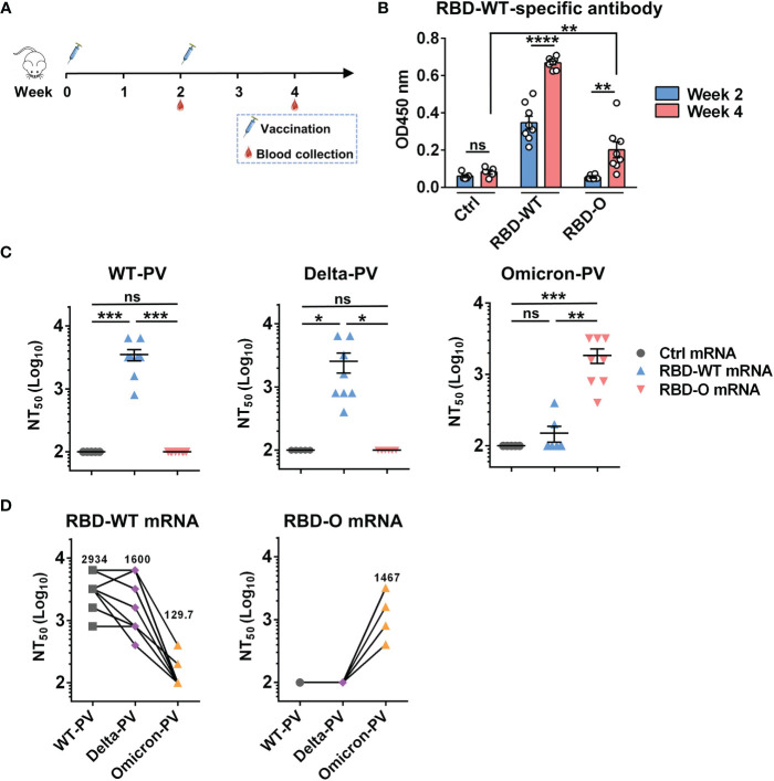
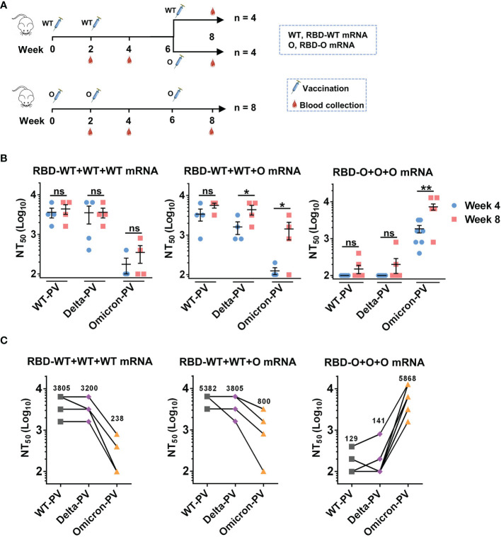
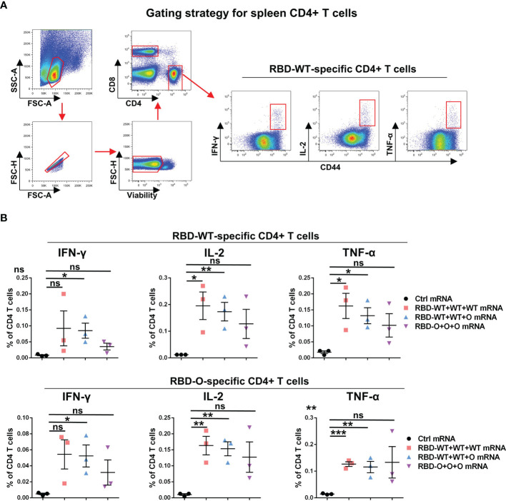
The newly emerged Omicron variant of severe acute respiratory syndrome coronavirus 2 (SARS-CoV-2) contains more than 30 mutations on the spike protein, 15 of which are located within the receptor binding domain (RBD). Consequently, Omicron is able to extensively escape existing neutralizing antibodies and may therefore compromise the efficacy of current vaccines based on the original strain, highlighting the importance and urgency of developing effective vaccines against Omicron. Here we report the rapid generation and evaluation of an mRNA vaccine candidate specific to Omicron, and explore the feasibility of heterologous immunization with WT and Omicron RBD vaccines. This mRNA vaccine encodes the RBD of Omicron (designated as RBD-O) and is formulated with lipid nanoparticle. Two doses of the RBD-O mRNA vaccine efficiently induce neutralizing antibodies in mice; however, the antisera are effective only on the Omicron variant but not on the wildtype and Delta strains, indicating a narrow neutralization spectrum. It is noted that the neutralization profile of the RBD-O mRNA vaccine is opposite to that observed for the mRNA vaccine expressing the wildtype RBD (RBD-WT). Importantly, booster with RBD-O mRNA vaccine after two doses of RBD-WT mRNA vaccine can significantly increase neutralization titers against Omicron. Additionally, an obvious increase in IFN-γ, IL-2, and TNF-α-expressing RBD-specific CD4+ T cell responses was observed after immunization with the RBD-WT and/or RBD-O mRNA vaccine. Together, our work demonstrates the feasibility and potency of an RBD-based mRNA vaccine specific to Omicron, providing important information for further development of heterologous immunization program or bivalent/multivalent SARS-CoV-2 vaccines with broad-spectrum efficacy.
Keywords: SARS-CoV-2; mRNA vaccine; neutralizing antibody; omicron variant; receptor-binding domain.
Copyright © 2022 Zang, Yin, Xu, Qiao, Liu, Lavillette, Zhang, Wang and Huang.
Conflict of interest statement
The authors declare that the research was conducted in the absence of any commercial or financial relationships that could be construed as a potential conflict of interest.
Figures
Similar articles
-
A mosaic-type trimeric RBD-based COVID-19 vaccine candidate induces potent neutralization against Omicron and other SARS-CoV-2 variants.Elife. 2022 Aug 25;11:e78633. doi: 10.7554/eLife.78633. Elife. 2022. PMID: 36004719 Free PMC article.
-
Pan-beta-coronavirus subunit vaccine prevents SARS-CoV-2 Omicron, SARS-CoV, and MERS-CoV challenge.J Virol. 2024 Sep 17;98(9):e0037624. doi: 10.1128/jvi.00376-24. Epub 2024 Aug 27. J Virol. 2024. PMID: 39189731 Free PMC article.
-
A Glycosylated RBD Protein Induces Enhanced Neutralizing Antibodies against Omicron and Other Variants with Improved Protection against SARS-CoV-2 Infection.J Virol. 2022 Sep 14;96(17):e0011822. doi: 10.1128/jvi.00118-22. Epub 2022 Aug 16. J Virol. 2022. PMID: 35972290 Free PMC article.
-
Clinical development of variant-adapted BNT162b2 COVID-19 vaccines: the early Omicron era.Expert Rev Vaccines. 2023 Jan-Dec;22(1):650-661. doi: 10.1080/14760584.2023.2232851. Expert Rev Vaccines. 2023. PMID: 37417000 Review.
-
Efficacy of mRNA, adenoviral vector, and perfusion protein COVID-19 vaccines.Biomed Pharmacother. 2022 Feb;146:112527. doi: 10.1016/j.biopha.2021.112527. Epub 2021 Dec 10. Biomed Pharmacother. 2022. PMID: 34906769 Free PMC article. Review.
Cited by
-
Booster vaccination with Ad26.COV2.S or an Omicron-adapted vaccine in pre-immune hamsters protects against Omicron BA.2.NPJ Vaccines. 2023 Mar 16;8(1):40. doi: 10.1038/s41541-023-00633-x. NPJ Vaccines. 2023. PMID: 36927774 Free PMC article.
-
Lessons from SENCOVAC: a prospective study evaluating the response to SARS-CoV-2 vaccination in the CKD spectrum.Nefrologia. 2022 Dec 16. doi: 10.1016/j.nefro.2022.12.006. Online ahead of print. Nefrologia. 2022. PMID: 36540904 Free PMC article. Review.
-
A Marek's Disease Virus Messenger RNA-Based Vaccine Modulates Local and Systemic Immune Responses in Chickens.Viruses. 2024 Jul 18;16(7):1156. doi: 10.3390/v16071156. Viruses. 2024. PMID: 39066318 Free PMC article.
-
Lipid nanoparticles as adjuvant of norovirus VLP vaccine augment cellular and humoral immune responses in a TLR9- and type I IFN-dependent pathway.J Virol. 2024 Dec 17;98(12):e0169924. doi: 10.1128/jvi.01699-24. Epub 2024 Nov 4. J Virol. 2024. PMID: 39494905 Free PMC article.
-
Do We Really Need Omicron Spike-Based Updated COVID-19 Vaccines? Evidence and Pipeline.Viruses. 2022 Nov 10;14(11):2488. doi: 10.3390/v14112488. Viruses. 2022. PMID: 36366586 Free PMC article. Review.
References
Publication types
MeSH terms
Substances
Supplementary concepts
LinkOut - more resources
Full Text Sources
Other Literature Sources
Medical
Research Materials
Miscellaneous