. 2022 Jun 30:10:937180.
doi: 10.3389/fchem.2022.937180. eCollection 2022.

A Combination of Membrane Filtration and Raman-Active DNA Ligand Greatly Enhances Sensitivity of SERS-Based Aptasensors for Influenza A Virus

Affiliations

A Combination of Membrane Filtration and Raman-Active DNA Ligand Greatly Enhances Sensitivity of SERS-Based Aptasensors for Influenza A Virus

Gleb Zhdanov et al. Front Chem. .

Abstract

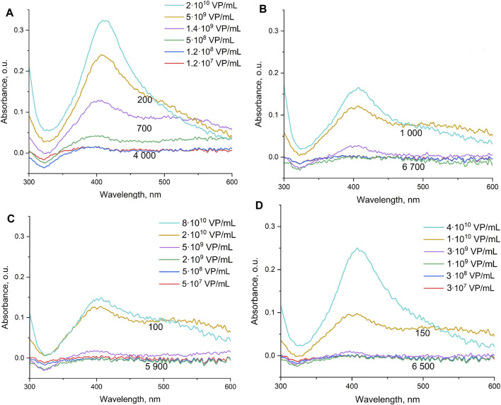
Biosensors combining the ultrahigh sensitivity of surface-enhanced Raman scattering (SERS) and the specificity of nucleic acid aptamers have recently drawn attention in the detection of respiratory viruses. The most sensitive SERS-based aptasensors allow determining as low as 104 virus particles per mL that is 100-fold lower than any antibody-based lateral flow tests but 10-100-times higher than a routine polymerase chain reaction with reversed transcription (RT-PCR). Sensitivity of RT-PCR has not been achieved in SERS-based aptasensors despite the usage of sophisticated SERS-active substrates. Here, we proposed a novel design of a SERS-based aptasensor with the limit of detection of just 103 particles per ml of the influenza A virus that approaches closely to RT-PCR sensitivity. The sensor utilizes silver nanoparticles with the simplest preparation instead of sophisticated SERS-active surfaces. The analytical signal is provided by a unique Raman-active dye that competes with the virus for the binding to the G-quadruplex core of the aptamer. The aptasensor functions even with aliquots of the biological fluids due to separation of the off-target molecules by pre-filtration through a polymeric membrane. The aptasensor detects influenza viruses in the range of 1·103-5·1010 virus particles per ml.

Keywords: SERS; aptamer; colloidal nanoparticle; influenza; membrane; sensor; virus.


Conflict of interest statement

The authors declare that the research was conducted in the absence of any commercial or financial relationships that could be construed as a potential conflict of interest.

Figures

FIGURE 1
FIGURE 1
Interaction between the aptamer and viruses assessed by biolayer interferometry. (A) Specificity of the aptamer RHA0385 toward the influenza A virus (IvA) compared to the Newcastle disease virus (NDV), influenza B virus (IvB), or virus-free allantoic fluid. The experiments were performed in the buffer with Tween-20 and bovine serum albumin to eliminate non-specific interactions. (B) Visualization of an assembly of sandwich-like complexes in the buffer without Tween-20 and bovine serum albumin. The aptamer-functionalized sensor interacts with 1) 4·103 HAU/ml of IvA; 2) aptamer-functionalized silver nanoparticles; 3) 4·103 HAU/ml of IvA with aggregated aptamer-functionalized silver nanoparticles; 4) 4·103 HAU/ml of IvA with a soluble aptamer; and 5) first, with 4·103 HAU/ml of IvA and second, with aptamer-functionalized silver nanoparticles.
FIGURE 2
FIGURE 2
Color changes in the concentrated solutions of viruses. IvA–influenza A virus—from left to right—2·1010 VP/ml; 5·109 VP/ml; 1.4·109 VP/ml; 5·108 VP/ml; 1.2·108 VP/ml; and 1.2 107 VP/ml. AF–allantoic fluid–dilutions are the same as for the influenza A virus. IvB–influenza B virus—from left to right—8·1010 VP/ml; 2·1010 VP/ml; 5·109 VP/ml; 2·109 VP/ml; 5·108 VP/ml; and 5·107 VP/ml; NDV–Newcastle disease—from left to right—4·1010 VP/ml; 1.0·1010 VP/ml; 3·109 VP/ml; 1·109 VP/ml; 2·108 VP/ml; and 2·107 VP/ml.
FIGURE 3
FIGURE 3
Spectra of samples with different quantities of the influenza A virus (A), allantoic fluid (dilutions are the same as for influenza A virus) (B), influenza B (C), and Newcastle disease virus (D). Numbers indicate the mean SERS intensity of BODIPY FL in the probes. The concentration dependencies were described earlier in Zhdanov et al. (2022).
FIGURE 4
FIGURE 4
Fraction of the aggregates of silver nanoparticles below 100 nm depending on the viral content. IvA–influenza A virus, IvB–influenza B virus, NDV–Newcastle disease virus, and AF–allantoic fluid. The dilution of allantoic fluid corresponds to the dilution of IvA.
FIGURE 5
FIGURE 5
Binding of the BHQ-2 amine to the aptamer RHA0385 estimated by biolayer interferometry. The experiment was performed in (A) a buffer with physiological ionic strength (buffer A) and in (B) a 50x-diluted buffer (buffer B).
FIGURE 6
FIGURE 6
SERS spectra from the BHQ-2 amine in the concentration of 8 nM: (A) on aggregated aptamer-functionalized silver nanoparticles in buffer C and in the presence of the Newcastle disease virus (NDV, 1011 VP/ml in allantoic fluid); (B) on aggregated silver nanoparticles and aggregated aptamer-functionalized silver nanoparticles; green line indicates the Raman spectrum of the BHQ-2 amine in the absence of nanoparticles.
FIGURE 7
FIGURE 7
Interaction between the aptamer and its complex with the BHQ-2 amine and the influenza A virus assessed by biolayer interferometry. The aptamer was incubated in the buffer or in a 1 µM solution of the BHQ-2 amine and then interacts with 4·103 HAU/ml solution of IvA.
FIGURE 8
FIGURE 8
Scanning electron microscopy images of the polyethylene terephthalate membrane with a pore size of 320 nm. (A) Front view; (B) side view.
FIGURE 9
FIGURE 9
Atomic force microscopy images of the influenza A viruses that passed the membrane (A) and were retained by the membrane (B). The maximum height on the A image is in the rate of 30–40 nm; on the B image, it is nearly 180–200 nm.
FIGURE 10
FIGURE 10
Schematic representation of the proposed sensor. Small objects are filtered out, and large objects displace the BHQ-2 amine from the complex with the aptamer decreasing the SERS signal in the probe.
FIGURE 11
FIGURE 11
SERS-based aptasensor for influenza A virus. (A) Absolute SERS signals for different dilutions of the influenza A virus, influenza B virus, or allantoic fluid. Dilution 100 corresponds to the undiluted sample. The experiments performed simultaneously are shown with the same symbols but in different colors. (B) Relative SERS signals obtained by dividing the SERS signal in the control experiment by the SERS signal in the sample with the influenza A virus. Setups with and without membranes are compared. Arrows indicate the limits of detection for each setup. The relative mean standard deviation for the measurement was 15%; thus, the samples with the ratio in the range 0.85–1.15 were interpreted as non-detectable viruses.

Similar articles

Cited by

References

    1. Akhmetova A. I., Yaminsky I. V. (2022). High Resolution Imaging of Viruses: Scanning Probe Microscopy and Related Techniques. Methods 197, 30–38. 10.1016/j.ymeth.2021.06.011 - DOI - PubMed
    1. Ambartsumyan O., Gribanyov D., Kukushkin V., Kopylov A., Zavyalova E. (2020). SERS-based Biosensors for Virus Determination with Oligonucleotides as Recognition Elements. Int. J. Mol. Sci. 21, 3373. 10.3390/ijms21093373 - DOI - PMC - PubMed
    1. Apel P. Y. (1995). Heavy Particle Tracks in Polymers and Polymeric Track Membranes. Radiat. Meas. 25, 667–674. 10.1016/1350-4487(95)00219-5 - DOI
    1. Asiala S. M., Marr J. M., Gervinskas G., Juodkazis S., Schultz Z. D. (2015). Plasmonic Color Analysis of Ag-Coated Black-Si SERS Substrate. Phys. Chem. Chem. Phys. 17, 30461–30467. 10.1039/C5CP04506A - DOI - PMC - PubMed
    1. Bizyaeva A. A., Bunin D. A., Moiseenko V. L., Gambaryan A. S., Balk S., Tashlitsky V. N., et al. (2021). The Functional Role of Loops and Flanking Sequences of G-Quadruplex Aptamer to the Hemagglutinin of Influenza a Virus. Int. J. Mol. Sci. 22, 2409. 10.3390/ijms22052409 - DOI - PMC - PubMed