

A Novel Calcium Phosphate-Based Nanocomposite for Augmentation of Cortical Bone Trajectory Screw Fixation
- PMID: 35844971
- PMCID: PMC9278980
- DOI: 10.2147/IJN.S365149
A Novel Calcium Phosphate-Based Nanocomposite for Augmentation of Cortical Bone Trajectory Screw Fixation
Abstract
Purpose: To evaluate the effect of cement augmentation of cortical bone trajectory (CBT) screws using a novel calcium phosphate-based nanocomposite (CPN).
Material and methods: CBT screws were placed into cadaveric lumbar vertebrae. Depending on the material used for augmentation, they were divided into the following three groups: CPN, polymethylmethacrylate (PMMA), and control. Radiological imaging was used to evaluate the cement dispersion. Biomechanical tests were conducted to measure the stability of CBT screws. A rat cranial defect model was used to evaluate biodegradation and osseointegration of the CPN.
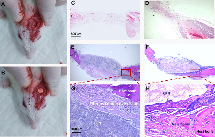
Results: After cement augmentation, the CPN tended to disperse into the distal part of the screws, whereas PMMA remained limited to the proximal part of the screws (P < 0.05). As for cement morphology, the CPN tended to form a concentrated mass, whereas PMMA arranged itself as a scattered cement cloud, but the difference was not significant (P > 0.05). The axial pullout test showed that the average maximal pullout force (Fmax) of CPN-augmented CBT screws was similar to that of the PMMA group (CPN, 1639.56 ± 358.21 N vs PMMA, 1778.45 ± 399.83 N; P = 0.745) and was significantly greater than that of the control group (1019.01 ± 371.98 N; P < 0.05). The average torque value in the CPN group was higher than that in the control group (CPN, 1.51 ± 0.78 N∙m vs control, 0.97 ± 0.58 N∙m) and lower than that in the PMMA group (1.93 ± 0.81 N∙m), but there were no statistically significant differences (P > 0.05). The CPN could be biodegraded and gradually replaced by newly formed bone tissue after 12 weeks in a rat cranial defect model.
Conclusion: The biocompatible CPN could be a valuable augmentation material to enhance CBT screw stability.
Keywords: CBT screws; CPN; PMMA; cement augmentation; osteoporotic spine.
© 2022 Wang et al.
Conflict of interest statement
Professor Lei Yang reports grants from NSFC, China, grants from MOST, China, during the conduct of the study. The authors report no conflicts of interest related to this work.
Figures


References
-
- Sakaura H, Miwa T, Yamashita T, Kuroda Y, Ohwada T. Posterior lumbar interbody fusion with cortical bone trajectory screw fixation versus posterior lumbar interbody fusion using traditional pedicle screw fixation for degenerative lumbar spondylolisthesis: a comparative study. J Neurosurg Spine. 2016;25(5):591–595. doi: 10.3171/2016.3.SPINE151525 - DOI - PubMed
-
- Crawford CH 3rd, Owens RK 2nd, Djurasovic M, Gum JL, Dimar JR 2nd, Carreon LY. Minimally-Invasive midline posterior interbody fusion with cortical bone trajectory screws compares favorably to traditional open transforaminal interbody fusion. Heliyon. 2019;5(9):e02423. doi: 10.1016/j.heliyon.2019.e02423 - DOI - PMC - PubMed
MeSH terms
Substances
LinkOut - more resources
Full Text Sources
Research Materials
