. 2022 Jul 11:15:3901-3923.
doi: 10.2147/JIR.S367193. eCollection 2022.

Characterization of Early Inflammatory Events Leading to Provoked Vulvodynia Development in Rats

Affiliations

Characterization of Early Inflammatory Events Leading to Provoked Vulvodynia Development in Rats

Yaseen Awad-Igbaria et al. J Inflamm Res. .

Abstract

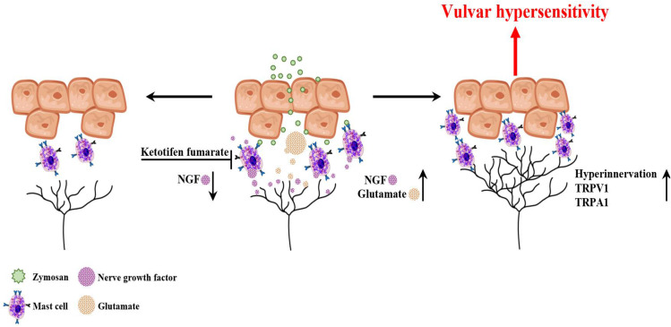
Background: Provoked vulvodynia (PV) is the main cause of vulvar pain and dyspareunia. The etiology of PV has not yet been elucidated. However, PV is associated with a history of recurrent inflammation, and its often accompanied by increases in the numbers of mast cells (MCs) and sensory hyperinnervation in the vulva. Therefore, this study aimed to examine the role of MCs and the early inflammatory events in the development of chronic vulvar pain in a rat model of PV.

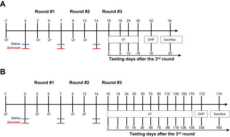
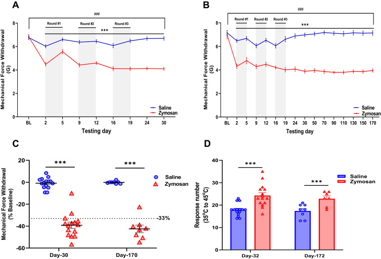
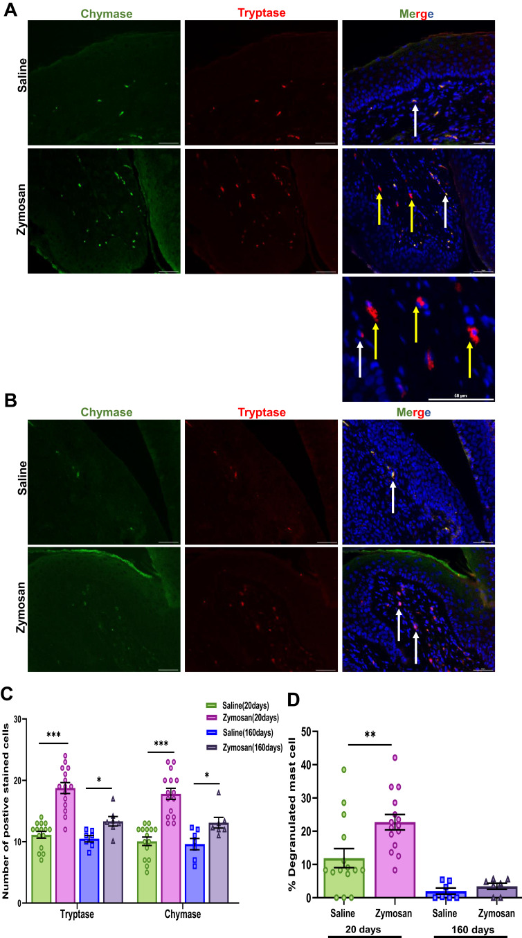
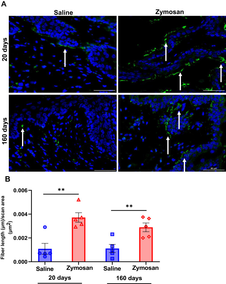
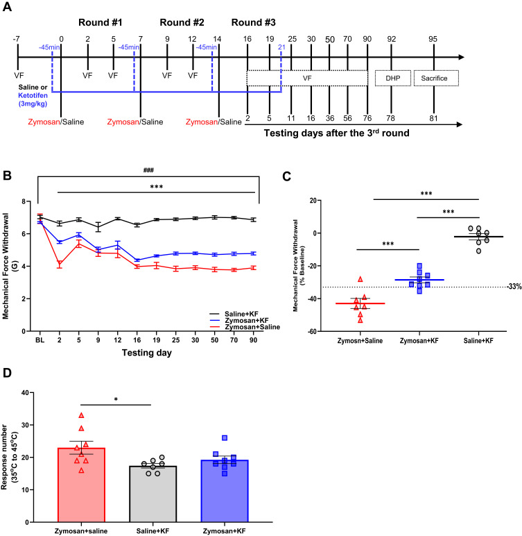
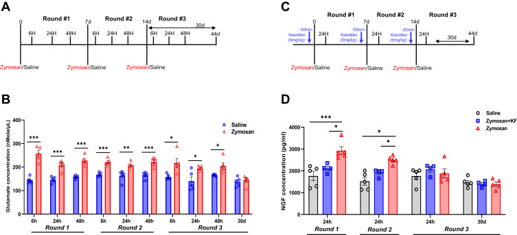
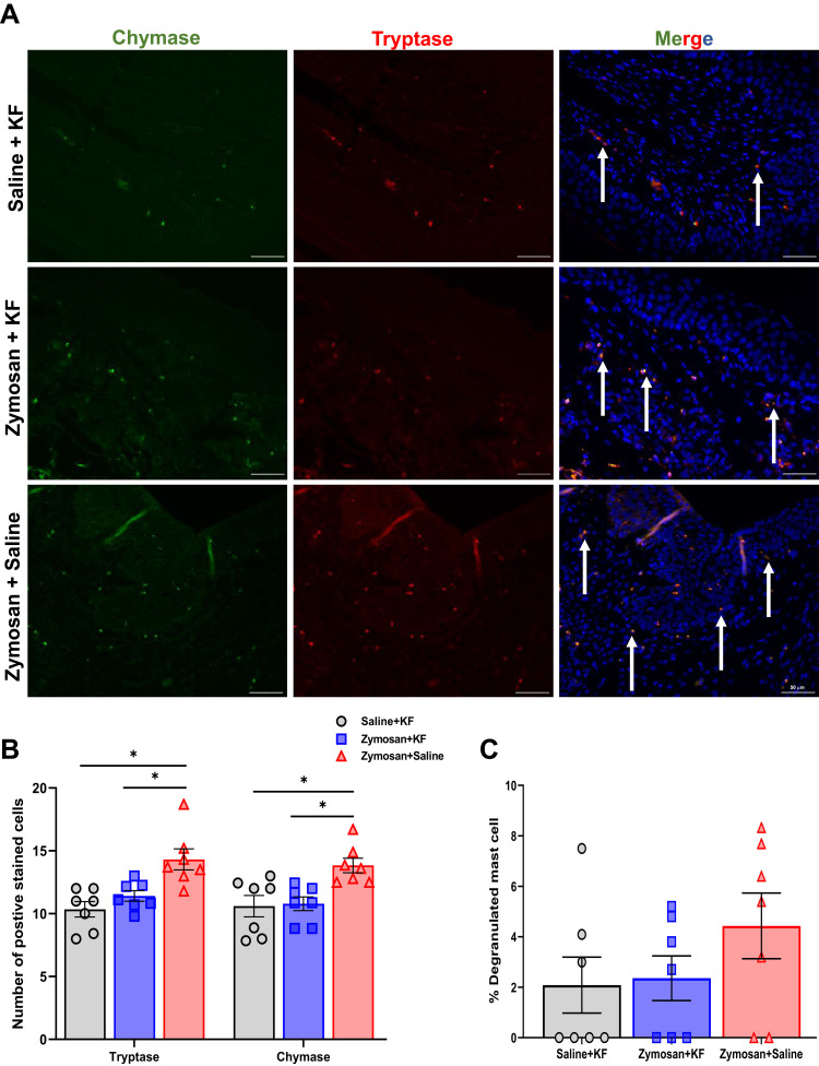
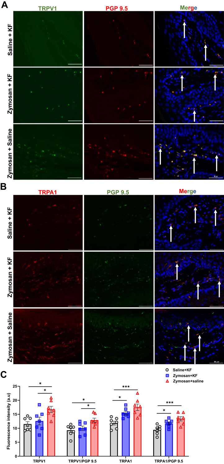
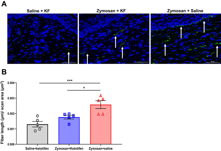
Methods: Mechanical and thermal vulvar sensitivity was measured for 5 months following zymosan vulvar challenges. Vulvar changes in glutamate and nerve growth factor (NGF) were analyzed using ELISA. Immunofluorescence (IF) staining of the vulvar section after 20, 81, and 160 days of the zymosan challenge were performed to test MCs accumulation, hyperinnervation, and expression of pain channels (transient receptor potential vanilloid/ankyrin-1-TRPV1 & TRPA1) in vulvar neurons. Changes in the development of vulvar pain were evaluated following the administration of the MCs stabilizer ketotifen fumarate (KF) during zymosan vulvar challenges.

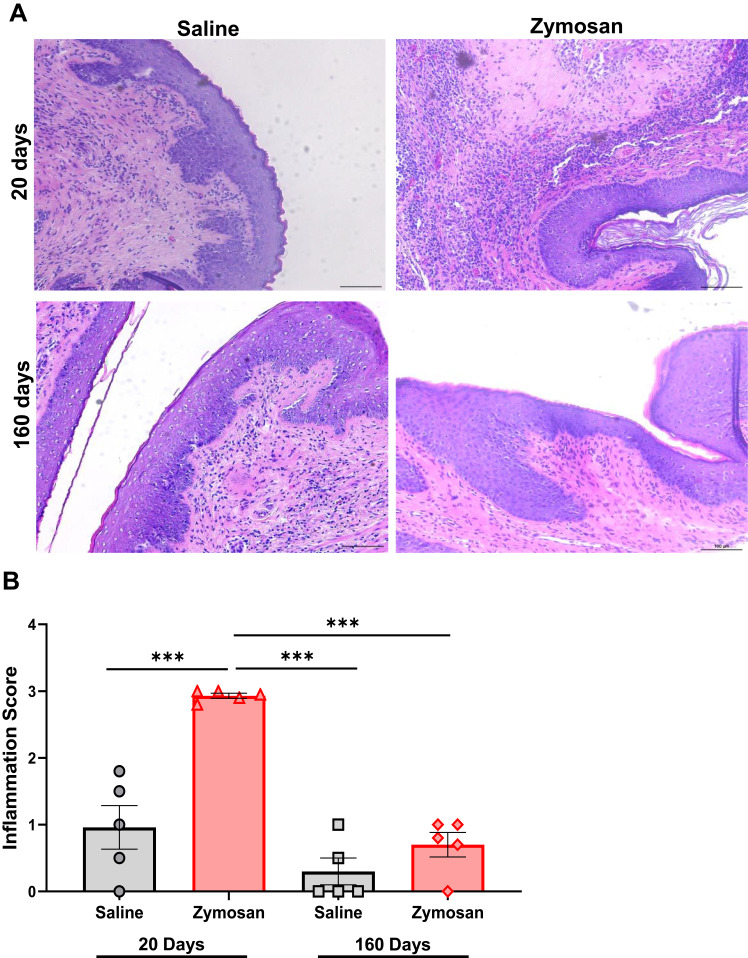
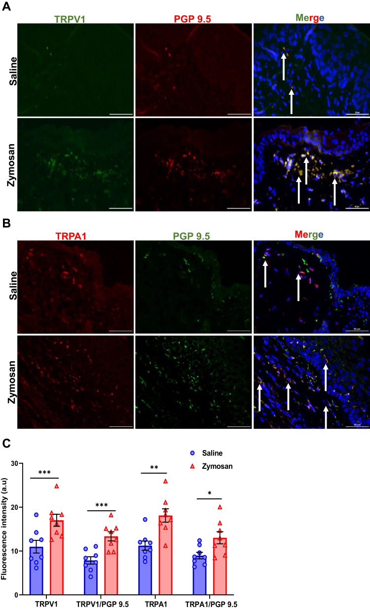
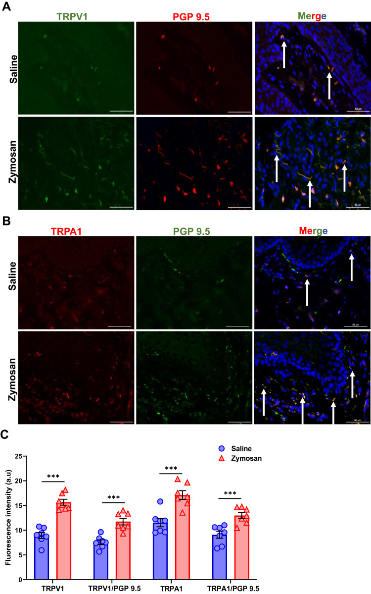
Results: Zymosan-challenged rats developed significant mechanical and thermal vulvar sensitivity that persisted for over 160 days after the zymosan challenge. During inflammation, increased local concentrations of NGF and glutamate and a robust increase in MCs degranulation were observed in zymosan-challenged rats. In addition, zymosan-challenged rats displayed sensory hyperinnervation and an increase in the expression of TRPV1 and TRPA1. Treatment with KF attenuated the upregulated level of NGF during inflammation, modulated the neuronal modifications, reduced MCs accumulation, and enhanced mechanical hypersensitivity after repeated inflammation challenges.

Conclusion: The present findings suggest that vulvar hypersensitivity is mediated by MCs accumulation, nerve growth, and neuromodulation of TRPV1 and TRPA1. Hence, KF treatment during the critical period of inflammation contributes to preventing chronic vulvar pain development.

Keywords: glutamate; hyperinnervation; inflammation; mast cell; nerve growth factor; provoked vulvodynia.


Conflict of interest statement

The authors declare that they have no conflicts of interest in relation to this work.

Figures

Figure 1
Figure 1
The experimental timeline illustrates the procedures across three rounds of vulvovaginal zymosan\saline administration. (A and B) Rats received zymosan\saline injections on days 0, 7, and 14; overall, three injections, 7 days between each one. Withdrawal responses of vulvar mechanical sensitivity were measured by electronic Von Frey (eVF) at baseline (−7 days) and +2 days and +5 days after zymosan\saline administration. eVF’s follow-up tests were taken at two points for the “short term“ effects of zymosan administration: day 24 (10 days after the 3rd round) and day 30 (16 days after the 3rd round). Thermal sensitivity was assessed using a Dynamic Hot plate test on day 32 (A). (B) for the ”long-term” effects of zymosan administration: eVF measurements were documented at nine points: day 24, day 30, day 50, day 70, day 90, day 110, day 130, day 150, day 170. Thermal allodynia was assessed using a Dynamic Hot plate test on day 172.
Figure 2
Figure 2
Development of vulvar mechanical and thermal sensitivity in rats after three rounds of zymosan administration. (A and B) Mechanical force withdrawal (G). There was a significant decrease in mechanical threshold among the saline group during each round. Afterward, the mechanical threshold returns to baseline ((A), short term n=15; (B), long term n=8). Among the zymosan group, the decrease in mechanical threshold was maintained throughout the zymosan challenges and 156 days after the 3rd round (day 170), compared to the baseline and saline group ((A), short term n=15; (B), long term n=8). (C) Mechanical force withdrawal (G) of day 30 and day 170 normalized to the baseline (D) Sum of nociceptive response (Vulva/hind paw licking, Rearing, and jumps) in the Dynamic Hot plate test (35°C to 45°C) after 18 days (day 32) and 158 days (day 172) of the 3rd round of zymosan/saline administration (n=8-15 per group). Mean ± SEM. ***P<0.001, zymosan vs saline, ###P<0.001, compared to Baseline.
Figure 3
Figure 3
Short and long-term effects of zymosan challenges in the vulva on mast cell. (A) Positively stained cells with mast cell chymase and tryptase in the zymosan and the saline group after 20 days of the 3rd round. Mast cell chymase (green; White arrows) and tryptase (red; white arrows), diffuse tryptase (Yellow arrows) merged with dapi stain (blue) scale bar: 50 µm. (B) Positively stained cells with mast cell chymase and tryptase in the zymosan and the saline group after 160 days of the 3rd round. Mast cell chymase (green; White arrows) and tryptase (red; white arrows) merged with dapi stain (blue) scale bar: 50 µm. (C) The number of positively chymase or tryptase stained cells in zymosan and saline groups after 20 and 160 days of the 3rd round (n=7-15 per group). (D) The percentage of degranulated mast cells among zymosan and saline groups after 20 and 160 days of the 3rd round (n=7-15 per group). Mean ± SEM. *P<0.05, **P<0.005, ***P<0.001.
Figure 4
Figure 4
Morphological and inflammatory effects of zymosan challenges in the vulva. (A) Visual inspection of hematoxylin and eosin (H&E)–stained sections, 20 and 160 days of zymosan/saline administration in the vulva. Saline administration in the vulva does not have morphological or inflammatory effects. However, evidence of an active inflammatory process, marked dermal lymphocytic infiltrate with exocytosis, increase in the number and the activation of mast cells (See Figure 3) after 20 days of the 3rd round of zymosan administration in the vulva. However, after 160 days of the 3rd round of zymosan administration, there was no evidence of inflammation. (B) Inflammation score of the saline and the zymosan groups after 20 and 160 of 3rd round of zymosan/saline administration (n=5 per group). Mean ± SEM. ***P<0.001.
Figure 5
Figure 5
TRPV1 and TRPA1 expression in vulvar nerves after multiple rounds of zymosan challenge. (A) The expression of TRPV1 channel in vulva nerves, 20 days after the 3rd zymosan/saline administration. Coexpression of TRPV1 channel (green) and neuronal PGP 9.5 (red; White arrows) merged with dapi stain (blue). (B) The expression of TRPA1 channel in vulva nerves, 20 days after the 3rd round zymosan\saline administration. Coexpression of TRPA1 channel (red) and neuronal PGP 9.5 (green; White arrows) merged with dapi stain (blue). Scale bar: 50 µm. (C) Fluorescence intensity (arbitrary units) of TRPV1, TRPA1, TRPV1/PGP-9.5, and TRPA1/PGP-9.5 in the zymosan and the saline groups 20 days of the 3rd round (n=8 per group). Mean ± SEM. *P<0.05, **P<0.005, ***P<0.001.
Figure 6
Figure 6
TRPV1 and TRPA1 expression in vulvar nerves after 160 days of the 3rd round of zymosan/saline administration. (A) The expression of TRPV1 channel in vulva nerves, 20 days after the 3rd zymosan/saline administration. Coexpression of TRPV1 channel (green) and neuronal PGP 9.5 (red; White arrows) merged with dapi stain (blue). (B) The expression of TRPA1 channel in vulva nerves, 20 days after the 3rd round zymosan\saline administration. Coexpression of TRPA1 channel (red) and neuronal PGP 9.5 (green; White arrows) merged with dapi stain (blue). Scale bar: 50 µm. (C) Fluorescence intensity (arbitrary units) of TRPV1, TRPA1, TRPV1/PGP-9.5, and TRPA1/PGP-9.5 in the zymosan and the saline groups 160 days of the 3rd round (n=8 per group). Mean ± SEM. ***P<0.001.
Figure 7
Figure 7
Hyperinnervation of vulvar nerves after multiple rounds of zymosan administration. (A) Nerve fibers [as detected by immunoreactivity (IR) for the pan-axonal marker protein gene product 9.5 (PGP 9.5; green)] merged with dapi stain (blue). Scale bar: 50 µm. (B) Fiber length (µm) per unit area (µm2) in the zymosan and saline groups after 20 and 160 days of the 3rd round of zymosan/saline administration (n=5 per group). Mean ± SEM. **P<0.005.
Figure 8
Figure 8
Treatment with Ketotifen fumarate during inflammation suppressed the development of vulvar hypersensitivity. (A) The experimental timeline illustrates the experimental procedures across three rounds of vulvar inflammation. Vulvar rat injected with zymosan or saline (n=7-8 per group) on days 0, 7, and 14; overall, three injections, 7 days between each one. The groups were injected with ketotifen (i.p; 3mg/Kg) or saline (i.p; 300ul) 45 minutes before the zymosan/saline administration into the vulva, and every day to day 7 after the 3rd round, overall 21 days of treatment. Vulvar mechanical sensitivity was measured by Von Frey (VF). Thermal sensitivity was assessed using a Dynamic Hot plate test on day 92 after the 3rd round. (B) Mechanical force withdrawal (G) of the zymosan-saline, the zymosan-ketotifen, and the saline-ketotifen group. There was no significant change in mechanical threshold among the saline- ketotifen group. There was a significant decrease in mechanical threshold in zymosan-saline and zymosan-ketotifen groups during the three rounds compared to the baseline. The decrease in mechanical threshold was maintained 76 days after the 3rd round in both groups. Also, there was a significant difference in mechanical force withdrawal between the two zymosan groups and the saline-ketotifen group at each time point (Excluding baseline). (C) Mechanical force withdrawal test of day 90 normalized to the baseline (n=7-8 per group). (D) Sum of nociceptive response in the Dynamic Hot plate test (35°C to 45°C) in zymosan-saline, zymosan-ketotifen, and saline-ketotifen group after 78 of the 3rd round of zymosan/saline administration (n=8 per group). Mean ± SEM. *P<0.05, ***P<0.001, difference between groups, ###P<0.001, Compared to the baseline.
Figure 9
Figure 9
Glutamate and NGF concentration in vulva tissue after zymosan or saline administration. (A) Rats received zymosan\saline injection (in the vulva) on days 0, 7, and 14; overall, three injections, 7 days between each one. Rats were sacrificed after 6, 24, and 48 hours of each injection and after 30 days of the 3rd injection of zymosan/saline. (B) Glutamate concentration in the vulva tissue of the saline and the zymosan group (n=5 per time point). (C) Rats were injected with ketotifen (i.p; 3mg/kg) 45 minutes before each zymosan/saline administration (in the vulva); overall, three injections of zymosan/saline, 7 days between each one. Rats were sacrificed after 24 hours of each injection and after 30 days of the 3rd injection of zymosan/saline. (D) NGF concentration in the vulva tissue of the saline, zymosan-saline, and zymosan-ketotifen group (n=4-5 per group at each time point). Mean ± SEM. *P <0.05, **P<0.005, ***P<0.001.
Figure 10
Figure 10
Long-term effects of mast cells stabilizer KF on the presence of mast cell, 81 days after the 3rd round. (A) Positively stained cells with mast cell chymase (green; white arrows) and tryptase (red; white arrows) merged with dapi stain (blue) in the Saline- ketotifen, the zymosan- ketotifen, and the zymosan-saline group. Scale bar: 50 µm. (B) The number of positively stained cells with chymase or tryptase in the Saline- ketotifen, the zymosan- ketotifen, and the zymosan-saline group after 81 days of the 3rd round (n=7 per group). (C) The percentage of degranulated mast cells among the Saline- ketotifen, the zymosan- ketotifen, and the zymosan-saline group after 81 days of the 3rd round (n=7 per group). Mean ± SEM. *P<0.05.
Figure 11
Figure 11
Long-term effects of mast cells stabilizer KF on TRPV1 and TRPA1 expression in vulvar nerves after 81 days of the 3rd round of zymosan/saline administration. (A) The expression of TRPV1 channel in vulva nerves, 81 days after the 3rd zymosan/saline administration. Coexpression of TRPV1 channel (green) and neuronal PGP 9.5 (red; white arrows) merged with dapi stain (blue). (B) The expression of TRPA1 channel in vulva nerves, 81 days after the 3rd round zymosan\saline administration. Coexpression of TRPA1 channel (red) and neuronal PGP 9.5 (green; white arrows) merged with dapi stain (blue). Scale bar: 50 µm. (C) Fluorescence intensity (arbitrary units) of TRPV1, TRPA1, TRPV1/PGP-9.5, and TRPA1/PGP-9.5 in the Saline- ketotifen, the zymosan- ketotifen, and the zymosan-saline group 81 of the 3rd round (n=7 per group). Mean ± SEM. *P<0.05, ***P<0.001.
Figure 12
Figure 12
Long-term effects of mast cells stabilizer KF on vulvar hyperinnervation. (A) Nerve fibers [as detected by immunoreactivity (IR) for the pan-axonal marker protein gene product 9.5 (PGP 9.5; green)] merged with dapi stain (blue). Scale bar: 50 µm. (B) Fiber length (µm) per unit area (µm2) in the saline- ketotifen, the zymosan-Ketotifen, and the zymosan-saline group (n=5 per group). Mean ± SEM. *P<0.05, ***P<0.001.
Figure 13
Figure 13
Diagram illustrating the involvement of mast cells in chronic vulvar pain development. Inflammation induced by zymosan leads to glutamate and NGF release from the inflammation area. The early inflammatory events (MC degranulation, NGF, and glutamate release) set the stage for long-term MC accumulation, nerves neuromodulation, and hyperinnervation manifested as vulvar hypersensitivity. In turn, mast cell stabilization by ketotifen fumarate attenuates the upregulated level of NGF during inflammation, modulates the neuronal modifications, reduces mast cells accumulation, and enhances mechanical hypersensitivity after repeated inflammation challenges.

Similar articles

Cited by

References

    1. Bornstein J, Goldstein AT, Stockdale CK, et al. 2015 ISSVD, ISSWSH and IPPS consensus terminology and classification of persistent vulvar pain and vulvodynia. Obstet Gynecol. 2016;127(4):745–751. doi:10.1097/AOG.0000000000001359 - DOI - PubMed
    1. Woolf CJ, Ma Q. Nociceptors—noxious stimulus detectors. Neuron. 2007;55(3):353–364. doi:10.1016/j.neuron.2007.07.016 - DOI - PubMed
    1. Bao C, Noga H, Allaire C, et al. Provoked vestibulodynia in women with pelvic pain. Sex Med. 2019;7(2):227–234. doi:10.1016/j.esxm.2019.03.002 - DOI - PMC - PubMed
    1. Henzell H, Berzins K, Langford JP. Provoked vestibulodynia: current perspectives. Int J Women's Health. 2017;9:631–642. doi:10.2147/IJWH.S113416 - DOI - PMC - PubMed
    1. Akopians AL, Rapkin AJ. Vulvodynia: the role of inflammation in the etiology of localized provoked pain of the vulvar vestibule (vestibulodynia). Semin Reprod Med. 2015;33(4):239–245. doi:10.1055/s-0035-1554919 - DOI - PubMed