Case Reports
. 2022 Jul 7:27:101653.
doi: 10.1016/j.ajoc.2022.101653. eCollection 2022 Sep.

Bacillary layer detachment in serpiginous-like choroiditis of presumed intraocular tuberculosis: Report of two cases

Affiliations
Case Reports

Bacillary layer detachment in serpiginous-like choroiditis of presumed intraocular tuberculosis: Report of two cases

Daniella Socci da Costa et al. Am J Ophthalmol Case Rep. .

Abstract

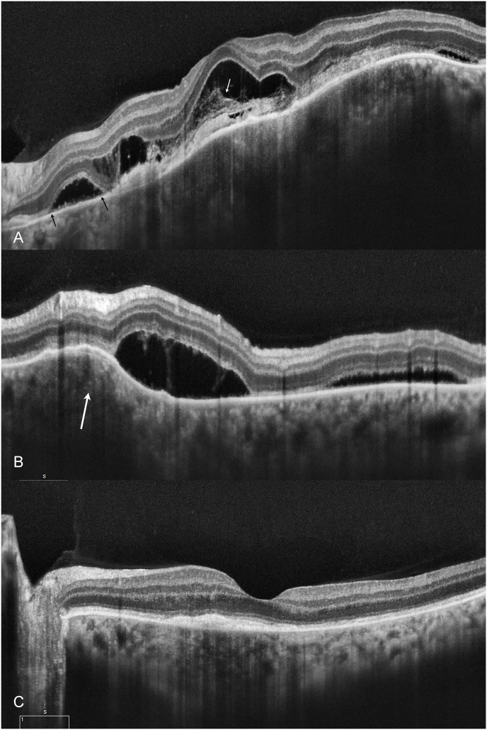
Purpose: To describe the presence of bacillary layer detachment (BALAD) in serpiginous-like choroiditis (SLC) in presumed intraocular tuberculosis.

Observations: Clinical and multimodal imaging including fundus photography, fundus autofluorescence, and spectral domain and enhanced-deep imaging optical coherence tomography (OCT) of two cases of SLC in presumed intraocular tuberculosis. Two patients (26 and 38-year-old woman) presented with unilateral, decreased vision of acute onset. They were diagnosed with SLC in presumed intraocular tuberculosis, and OCT revealed splitting of the ellipsoid zone, resembling BALAD. All two patients showed complete resolution after treatment with antitubercular therapy (ATT).

Conclusions and importance: BALAD appears in the acute stage of SLC in presumed intraocular tuberculosis and resolves rapidly at the beginning of ATT.

Keywords: Bacillary layer detachment; Optical coherence tomography; Photoreceptor layer; Serpiginous-like choroiditis; Tuberculosis; Uveitis.


Conflict of interest statement

The following authors have no financial disclosures: da Costa D.S.; Silva A. G., Melichar A., Neves D. B., Correa P. A., Moraes R. T.

Figures

Fig. 1
Fig. 1
(A) Ultra-wide-field fundus retinography of the left eye of the patient in case 1, at presentation showing multiple deep ill-defined yellowish lesions, from the optic disc toward the posterior pole and nasal retina (B) Ultra-wide-field autofluorescence showing multiple hyperfluorescent diffuse lesions, corresponding active lesions. (C, D) Retinography after 1 month without treatment showing more active lesions. (E) The pigmented lesions after 2 months of treatment. (F) The lesions show total hypoautofluorescence after healing.
Fig. 2
Fig. 2
Ultra-wide-field fluorescein angiography at presentation. In the early phase (A) the multifocal lesions show early hypofluorescence with late hyperfluorescence (B) indicating active choroidal inflammation.
Fig. 3
Fig. 3
Optical coherence tomography at presentation. (A) In the scan at the nasal retina, there is presence of BALAD (white asterisk). The outer retina split occurs at the level of the hyporreflective myoid zone (MZ) leaving the remaining photoreceptor layers adherent to subretinal hyperreflective material anterior to the retinal pigment epithelium (RPE)-basal lamina-Bruch membrane complex at the base of the BALAD. A choroidal lesion is causing an elevation of the retina (blue arrow). (B) A dome-shaped cystic retinal hyporreflective space superiorly to the macula. (C) Subretinal fluid accumulation is observed inferiorly to the macula. (D) Optical coherence tomography at 2 months of follow-up, shows a resolution of the BALAD and a hyperreflective material is noted over the RPE. (For interpretation of the references to colour in this figure legend, the reader is referred to the Web version of this article.)
Fig. 4
Fig. 4
Confocal digital fundus retinography of the left eye of patient 2. (A) At presentation, the fundus photograph showing deep ill-defined multifocal and confluent yellowish lesions involving the macula. A choroidal lesion is causing an elevation of the retina (white arrow) (B) and (C) at 7 and 14-day follow-up, respectively. The border of the lesions becomes progressively more distinct, and an amoeboid-like pattern can be noted (arrows). (D) At 45-day follow-up the healing of these lesions is evident. Irregular retina pigment epithelium (RPE) perturbations, diffuse RPE mottling with extensive atrophy of RPE and choriocapillaris are observed.
Fig. 5
Fig. 5
(A and B). Optical coherence tomography at presentation (scan sites are the solid and dashed lines in Fig. 1A, respectively). (A) A dome-shaped cystic retinal hyporreflective space. A thin line of reflectivity at the base of the cystic space appears continuous with the flanking ellipsoid zone (black arrow). In the subfoveal area, the retinal cavity contains an amorphous hyperreflective material along the outer retinal surface (white arrow). (B) Subretinal fluid accumulation is observed inferiorly to the fovea. A choroidal lesion is causes retina elevation (white arrow). (C) Optical coherence tomography at the 45-day follow-up. A subretinal hyperreflective material is noted under the fovea.
Fig. 6
Fig. 6
(A and B). Fluorescein angiography at presentation. In the early phase (A) the macular lesions show early hypofluorescence. In the late phase (B), the lesions become progressively hyperfluorescent. (C) Ultra-wide-field autofluorescence at the 7-day follow-up. The central inactive lesions present a hyperfluorescent amoeboid-like pattern. They are surrounded by a hypofluorescent area, corresponding to active lesions (black arrows). There are peripheral patches of hyperfluorescence in a pattern mimicking multiple evanescent white dot syndrome (white arrows). (D) Ultra-wide-field autofluorescence at the 45-day follow-up. The areas of macular hyperfluorescence corresponding to healed lesions are observed. No peripheral lesions are noted.

References

    1. WHO . World Health Organization; Geneva: 2020. Global Tuberculosis Report 2020. Licence: CC BY-NC-SA 3.0 IGO.
    1. Gupta A., Bansal R., Gupta V., Sharma A., Bambery P. Ocular signs predictive of tubercular uveitis. Am J Ophthalmol. 2010;149:562–570. - PubMed
    1. Kawali A., Bavaharan B., Sanjay S., et al. Serpiginous-like choroiditis (SLC) –Morphology and treatment outcomes. Ocul Immunol Inflamm. 2019:1–9. 0(0) - PubMed
    1. Kohli G.M., Bhatia P., Sen A., Gupta A. Bacillary layer detachment in hyper-acute stage of acute posterior multifocal placoid pigment epitheliopathy: a case series. Ocul Immunol Inflamm. 2020 Sep;23:1–4. - PubMed
    1. Cicinelli MV, Giuffré C, Marchese A, et al. The bacillary detachment in posterior segment ocular diseases. Am Acad Opthalmol. - PubMed

Publication types

LinkOut - more resources