

Identification of Prefrontal Cortex and Amygdala Expressed Genes Associated With Sevoflurane Anesthesia on Non-human Primate
- PMID: 35845920
- PMCID: PMC9286018
- DOI: 10.3389/fnint.2022.857349
Identification of Prefrontal Cortex and Amygdala Expressed Genes Associated With Sevoflurane Anesthesia on Non-human Primate
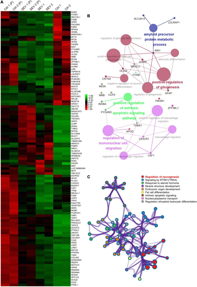
Abstract
Clinical trials and animal studies have indicated that long-term use or multiple administrations of anesthesia may lead to fine motor impairment in the developing brain. Most studies on anesthesia-induced neurotoxicity have focused on the hippocampus and prefrontal cortex (PFC); however, the role of other vital encephalic regions, such as the amygdala, is still unclear. Herein, we focused on sevoflurane, the most commonly used volatile anesthetic in infants, and performed a transcriptional analysis of the PFC and amygdala of macaques after multiple exposures to the anesthetic by RNA sequencing. The overall, overlapping, and encephalic region-specific transcriptional patterns were separately analyzed to reveal their functions and differentially expressed gene sets that were influenced by sevoflurane. Specifically, functional, protein-protein interaction, neighbor gene network, and gene set enrichment analyses were performed. Further, we built the basic molecular feature of the amygdala by comparing it to the PFC. In comparison with the amygdala's changing pattern following sevoflurane exposure, functional annotations of the PFC were more enriched in glial cell-related biological functions than in neuron and synapsis development. Taken together, transcriptional studies and bioinformatics analyses allow for an improved understanding of the primate PFC and amygdala.
Keywords: RNA sequencing; amygdala; prefrontal cortex; primate; sevoflurane.
Copyright © 2022 Cheng, Liu, Zhang and Jiang.
Conflict of interest statement
The authors declare that the research was conducted in the absence of any commercial or financial relationships that could be construed as a potential conflict of interest.
Figures


Similar articles
-
The effect of sevoflurane exposure on cell-type-specific changes in the prefrontal cortex in young mice.J Neurochem. 2024 Jun;168(6):1080-1096. doi: 10.1111/jnc.16068. Epub 2024 Feb 5. J Neurochem. 2024. PMID: 38317263
-
Epitranscriptomic Analysis of N6-methyladenosine in Infant Rhesus Macaques after Multiple Sevoflurane Anesthesia.Neuroscience. 2022 Feb 1;482:64-76. doi: 10.1016/j.neuroscience.2021.11.030. Epub 2021 Nov 27. Neuroscience. 2022. PMID: 34843896
-
Proteomic analysis of gene expression in the prefrontal cortex in infant rhesus macaques after multiple sevoflurane exposures.J Anesth. 2023 Dec;37(6):853-860. doi: 10.1007/s00540-023-03244-x. Epub 2023 Aug 22. J Anesth. 2023. PMID: 37608132
-
The developing amygdala: a student of the world and a teacher of the cortex.Curr Opin Psychol. 2017 Oct;17:55-60. doi: 10.1016/j.copsyc.2017.06.012. Epub 2017 Jun 23. Curr Opin Psychol. 2017. PMID: 28950973 Free PMC article. Review.
-
Anesthesia and the Developing Brain: A Review of Sevoflurane-induced Neurotoxicity in Pediatric Populations.Clin Ther. 2021 Apr;43(4):762-778. doi: 10.1016/j.clinthera.2021.01.024. Epub 2021 Mar 3. Clin Ther. 2021. PMID: 33674065 Review.
Cited by
-
General anesthetic action profile on the human prefrontal cortex cells through comprehensive single-cell RNA-seq analysis.iScience. 2023 Mar 31;26(4):106534. doi: 10.1016/j.isci.2023.106534. eCollection 2023 Apr 21. iScience. 2023. PMID: 37123239 Free PMC article.
References
LinkOut - more resources
Full Text Sources
Miscellaneous