

Value of Transabdominal Combined Transvaginal Color Doppler Ultrasonography in the Distinguish between Uterine Adenomyoma and Uterine Fibroids
- PMID: 35845931
- PMCID: PMC9283036
- DOI: 10.1155/2022/9599571
Value of Transabdominal Combined Transvaginal Color Doppler Ultrasonography in the Distinguish between Uterine Adenomyoma and Uterine Fibroids
Retraction in
-
Retracted: Value of Transabdominal Combined Transvaginal Color Doppler Ultrasonography in the Distinguish between Uterine Adenomyoma and Uterine Fibroids.Biomed Res Int. 2023 Nov 29;2023:9876769. doi: 10.1155/2023/9876769. eCollection 2023. Biomed Res Int. 2023. PMID: 38075366 Free PMC article.
Abstract
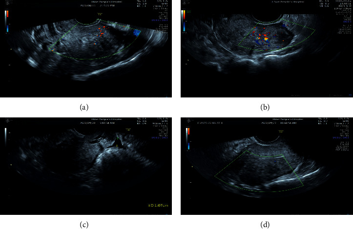
Objective: To investigate the value of transabdominal combined transvaginal color Doppler ultrasonography in the diagnosis of uterine adenomyoma.
Methods: A total of 80 patients with suspected uterine adenomyoma in our hospital from January 2019 to December 2021 were selected as the study subjects. All of them were examined by transabdominal color Doppler ultrasound (TA-CDUS) and transvaginal color Doppler ultrasound (TV-CDUS), and the postoperative pathological examination results were taken as the gold standard to analyze the diagnostic efficacy of different examination methods for uterine adenomyoma.
Results: By postoperative pathological biopsy, 46 cases (57.50%) were diagnosed as positive and 34 cases (42.50%) were diagnosed as negative, including 29 cases of uterine adenomyoma and 5 other cases. The sensitivity, accuracy, and negative predictive value of TA-CDUS combined with TV-CDUS in the diagnosis of adenomyoma were higher than those of TA-CDUS (P < 0.05), and the Kappa value between TA-CDUS and pathological diagnosis was 0.923, which was higher than the 0.615 between TV-CDUS and pathological diagnosis. TA-CDUS combined with TV-CDUS showed that there were significant differences in the distribution of Adier blood flow grades between patients with uterine adenomyoma and uterine fibroids (P < 0.05), and the Adier blood flow grades of patients with uterine adenomyoma were mainly grade 0 and grade I; and the resistance index (RI), peak systolic velocity (Vs), and pulsatile index (PI) in patients with uterine adenomyoma were higher than those in patients with uterine fibroids (P < 0.05).
Conclusion: Compared with TA-CDUS, TA-CDUS combined with TV-CDUS is more sensitive and accurate in the diagnosis of uterine adenomyoma and has a good consistency with pathological diagnosis results. Attention should be paid to the blood flow parameter values in the differential diagnosis of uterine fibroids.
Copyright © 2022 Hongmei Qi et al.
Conflict of interest statement
The authors declare no competing interests.
Figures
Similar articles
-
The Diagnostic Accuracy of Transabdominal and Transvaginal Color Doppler Ultrasound for Pregnant Women with Vasa Previa and Velamentous Cord Insertion.J Environ Public Health. 2022 Sep 29;2022:1685783. doi: 10.1155/2022/1685783. eCollection 2022. J Environ Public Health. 2022. Retraction in: J Environ Public Health. 2023 Sep 27;2023:9864294. doi: 10.1155/2023/9864294. PMID: 36213044 Free PMC article. Retracted.
-
Differentiation between adenomyoma and leiomyoma with transvaginal ultrasonography.Ultrasound Obstet Gynecol. 1995 Jan;5(1):47-50. doi: 10.1046/j.1469-0705.1995.05010047.x. Ultrasound Obstet Gynecol. 1995. PMID: 7850590 Clinical Trial.
-
Adenomyoma and leiomyoma: differential diagnosis with transvaginal sonography.J Clin Ultrasound. 1998 Jan;26(1):21-5. doi: 10.1002/(sici)1097-0096(199801)26:1<21::aid-jcu5>3.0.co;2-l. J Clin Ultrasound. 1998. PMID: 9475204
-
Primary ovarian adenomyoma in a woman with endometrial polyp: a case report and review of the literature.Arch Gynecol Obstet. 2009 Sep;280(3):445-8. doi: 10.1007/s00404-008-0913-z. Epub 2009 Jan 8. Arch Gynecol Obstet. 2009. PMID: 19130067 Review.
-
Flow characteristics in benign and malignant gynecologic tumors using transvaginal color flow Doppler.Obstet Gynecol. 1994 Jan;83(1):125-30. Obstet Gynecol. 1994. PMID: 8272293 Review.
Cited by
-
Retracted: Value of Transabdominal Combined Transvaginal Color Doppler Ultrasonography in the Distinguish between Uterine Adenomyoma and Uterine Fibroids.Biomed Res Int. 2023 Nov 29;2023:9876769. doi: 10.1155/2023/9876769. eCollection 2023. Biomed Res Int. 2023. PMID: 38075366 Free PMC article.
-
The Diagnostic Accuracy of Transabdominal and Transvaginal Color Doppler Ultrasound for Pregnant Women with Vasa Previa and Velamentous Cord Insertion.J Environ Public Health. 2022 Sep 29;2022:1685783. doi: 10.1155/2022/1685783. eCollection 2022. J Environ Public Health. 2022. Retraction in: J Environ Public Health. 2023 Sep 27;2023:9864294. doi: 10.1155/2023/9864294. PMID: 36213044 Free PMC article. Retracted.
-
Prolapsed Atypical Polypoid Adenomyoma-A Case Report and Literature Review.Life (Basel). 2023 Dec 15;13(12):2352. doi: 10.3390/life13122352. Life (Basel). 2023. PMID: 38137953 Free PMC article.
References
Publication types
MeSH terms
LinkOut - more resources
Full Text Sources
Research Materials
Miscellaneous
