. 2022 Jul 7:2022:1138709.
doi: 10.1155/2022/1138709. eCollection 2022.

Silencing lncRNA 93358 Inhibits the Apoptosis of Myocardial Cells in Myocardial Infarction Rats by Inducing the Expression of SLC8A1

Affiliations

Silencing lncRNA 93358 Inhibits the Apoptosis of Myocardial Cells in Myocardial Infarction Rats by Inducing the Expression of SLC8A1

Jiumei Cai et al. Biomed Res Int. .

Retraction in

Abstract

Objective: To explore the inhibitor effects and mechanism of lncRNA 93358 against the apoptosis of myocardial cells in rats with myocardial infarction.

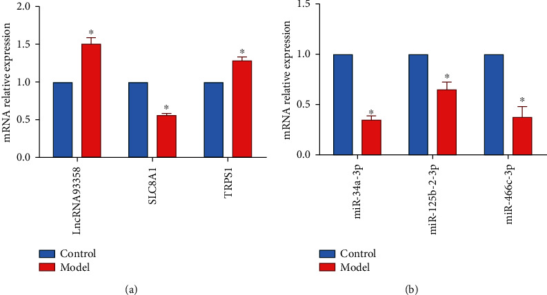
Methods: The myocardial infarction model was established in rats, which were identified by cardiac ultrasound. TTC staining was used to evaluate the degree of heart infarction, and HE staining was utilized to determine the pathological state in myocardial tissues. The apoptotic state in myocardial tissues was confirmed by TUNEL assay. lncRNA 93358 was screened out using a high-throughput sequencing which was confirmed by RT-qPCR. The interaction between miR-466c-3p and SLC8A1 was identified using the dual-luciferase reporter assay. The expression level of Bax, Bcl-2, and SLC8A1 was determined in lncRNA 93358 knockdown cells using RT-qPCR and Western blotting.

Results: Massive myocardial necrosis was observed in model rats according to the results of TTC staining, HE staining, and TUNEL assay. lncRNA 93358 and Bax were found significantly upregulated, and Bcl-2 and SLC8A1 were greatly downregulated in model rats, which were dramatically reversed by the knockdown of lncRNA 93358, accompanied by the decline area of myocardial necrosis and decreased apoptotic myocardial cells.

Conclusion: Silencing lncRNA 93358 inhibits the apoptosis of myocardial cells in rats with myocardial infarction by inducing the expression of SLC8A1.


Conflict of interest statement

The authors declare that they have no conflict of interest.

Figures

Figure 1
Figure 1
The pie chart of gene structure distribution. Percent of reads mapped to genome regions. M1-M3: model group; C1-C3: control group. Blue represents exons, orange represents introns, and green represents intergenic.
Figure 2
Figure 2
The line chart (a) and Venn diagram (b) of lncRNA concentrations.
Figure 3
Figure 3
Identification of differentially expressed lncRNA. (a) The PCA diagram. (b) Volcano map of differential expression analysis. The horizontal axis represented the expression multiple of the transcript between different groups of samples and the vertical axis represented the statistical significance of changes in transcript expression levels (q value). Red dots represented an upregulated transcript, green dots represented a downregulated transcript, and black represented a nondifferential transcript.
Figure 4
Figure 4
Functional enrichment of differentially expressed lncRNAs. (a) GO analysis on the differentially expressed lncRNAs. The horizontal axis represented a functional classification, and the vertical axis represented the number of genes in the classification (right) and their percentage in the total number of genes annotated (left). (b) KEGG analysis on the differentially expressed lncRNAs. The vertical axis represents the pathway category, and the vertical axis represents the number of genes in the classification (right) and its percentage in the total number of annotated genes (left). Different colors represent different categories.
Figure 5
Figure 5
Validation of sequencing data. (a and b) The expression levels of lncRNA 93358, SLC8A1, and TRPS1 (a) and miR-34a-3p, miR-125b-2-3p, and miR-466c-3p (b) in the rat myocardial tissues were detected by RT-qPCR. ∗∗P < 0.01.
Figure 6
Figure 6
The regulation axis of lncRNA 93358/miRNA/SLC8A1. (a and b) The expression of lncRNA 93358 and miR-466c-3p in the four rat myocardial tissues was measured by RT-qPCR. P < 0.05 vs. control, #P < 0.05 vs. model, and ^P < 0.05 vs. NC group. (c) The binding between SLC8A1 and miR-466c-3p was measured by dual-luciferase reporter. P < 0.05.
Figure 7
Figure 7
The results of cardiac ultrasound in rats. P < 0.05 vs. control, #P < 0.05 vs. model, and ^P < 0.05 vs. NC group. IVSd: interventricular septum size (diastole); IVSs: interventricular septum size (systole); LVIDd: left ventricular internal size (diastolic); LVIDs: internal size of left ventricle (systole); LVPWd: left ventricular posterior wall size (diastolic); LVPWs: posterior wall size of left ventricle (systole); EF: left ventricular ejection fraction (LVEF); FS: percentage of left ventricular fraction shortening; HR: heart rate; CO: cardiac output.
Figure 8
Figure 8
Pathological changes in the heart of rats in each group. (a) TTC staining of rat heart. (b) The images of HE staining.
Figure 9
Figure 9
Effect of lncRNA 93358 on apoptosis in myocardial infarction rat cardiomyocytes. (a) The apoptosis of myocardial tissues was determined by TUNEL assay. (b and c) The expression level of Bax, Bcl-2, and SLC8A1 in the myocardial cells was determined by RT-qPCR (b) and Western blotting (c). P < 0.05 vs. control, #P < 0.05 vs. model, and ^P < 0.05 vs. NC group.

References

    1. Jenča D., Melenovský V., Stehlik J., et al. Heart failure after myocardial infarction: incidence and predictors. ESC Heart Fail . 2021;8(1):222–237. doi: 10.1002/ehf2.13144. - DOI - PMC - PubMed
    1. Ruddox V., Sandven I., Munkhaugen J., Skattebu J., Edvardsen T., Otterstad J. E. Atrial fibrillation and the risk for myocardial infarction, all-cause mortality and heart failure: a systematic review and meta-analysis. European Journal of Preventive Cardiology . 2017;24(14):1555–1566. doi: 10.1177/2047487317715769. - DOI - PMC - PubMed
    1. Frangogiannis N. G. Pathophysiology of myocardial infarction. Comprehensive Physiology . 2015;5(4):1841–1875. doi: 10.1002/cphy.c150006. - DOI - PubMed
    1. Smit M., Coetzee A. R., Lochner A. The pathophysiology of myocardial ischemia and perioperative myocardial infarction. Journal of Cardiothoracic and Vascular Anesthesia . 2020;34(9):2501–2512. doi: 10.1053/j.jvca.2019.10.005. - DOI - PubMed
    1. Ghafouri-Fard S., Azimi T., Taheri M. Myocardial infarction associated transcript (MIAT): review of its impact in the tumorigenesis. Biomedicine & Pharmacotherapy . 2021;133:p. 111040. doi: 10.1016/j.biopha.2020.111040. - DOI - PubMed

Publication types

MeSH terms