. 2022 Jul 8:2022:7230167.
doi: 10.1155/2022/7230167. eCollection 2022.

Effect of MicroRNA-138 on Tumor Necrosis Factor-Alpha-Induced Suppression of Osteogenic Differentiation of Dental Pulp Stem Cells and Underlying Mechanism

Affiliations

Effect of MicroRNA-138 on Tumor Necrosis Factor-Alpha-Induced Suppression of Osteogenic Differentiation of Dental Pulp Stem Cells and Underlying Mechanism

Wenzhe Liu et al. Biomed Res Int. .

Abstract

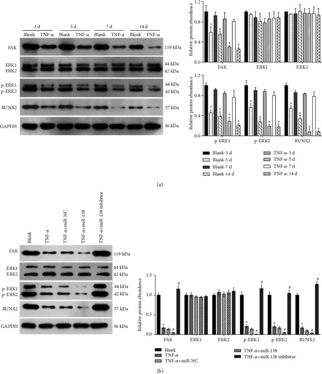
High doses of tumor necrosis factor-α (TNF-α) suppress osteogenic differentiation of human dental pulp stem cells (hDPSCs). In the present study, we aimed to explore the role and potential regulatory mechanism of microRNA-138 (miR-138) in the osteogenic differentiation of hDPSCs after treatment with a high dose of TNF-α. The hDPSCs were cultured in osteogenic medium with or without 50 ng/ml TNF-α. The miR-138 levels were upregulated during osteogenic differentiation of the hDPSCs following TNF-α treatment. The miR-138 overexpression accelerated but miR-138 knockdown alleviated the TNF-α-induced suppression of the alkaline phosphatase activity, calcium deposition, and protein abundance of dentin sialophosphoprotein, dentin matrix protein 1, bone sialoprotein, and osteopontin during osteogenic differentiation induction of hDPSCs. Additionally, miR-138 overexpression accelerated but miR-138 knockdown alleviated the suppression of the focal adhesion kinase- (FAK-) extracellular signal-regulated kinase 1/2 (ERK1/2) signaling pathway during osteogenic differentiation induction of hDPSCs under TNF-α treatment. In conclusion, miR-138 accelerates TNF-α-induced suppression of osteogenic differentiation of hDPSCs. Inactivation of the FAK-ERK1/2 signaling pathway may be one of the mechanisms underlying the effect of miR-138. Inhibition of miR-138 expression may be a strategy to weaken the inhibitory effect of high-dose TNF-α on the osteogenic differentiation of hDPSCs.


Conflict of interest statement

The authors declare no competing interests associated with the manuscript.

Figures

Figure 1
Figure 1
Expression of miR-138 cultured in osteogenic medium with or without treatment with 50 ng/mL TNF-α for 3, 5, 7, and 14 days, as detected via RT-qPCR. The bar graphs are means of relative miR-138 expression from three independent experiments. The error bars are standard deviations. P < 0.05, when compared to the without TNF-α group.
Figure 2
Figure 2
Isolation and culture of hDPSCs. (a) hDPSCs of first passage, second passage, and third passage were observed using an inverted microscope (magnification: 100x). (b) hDPSCs identified via flow cytometry analysis.
Figure 3
Figure 3
Effect of miR-138 on the osteogenic differentiation of hDPSCs under TNF-α treatment. hDPSCs were divided into 5 groups: the blank group (cultured in osteogenic medium), TNF-α (cultured in osteogenic medium with 50 ng/mL TNF-α), miR-NC + TNF-α group (pretransfected with miR-NC for 24 h and cultured in osteogenic medium with 50 ng/mL TNF-α), miR-138 + TNF-α group (pretransfected with miR-138 for 24 h and cultured in osteogenic medium with 50 ng/mL TNF-α), and miR-138 inhibitor + TNF-α group (pretransfected with miR-138 inhibitor for 24 h and cultured in osteogenic medium with 50 ng/mL TNF-α) and cultured for 7 days. After culturing for 7 days, cells were harvested for RT-qPCR and the detection of miR-138 expression levels (a), ALP activity assay (b), alizarin red S staining (c), and Western blotting to analyze the expression of DSPP, DMP-1, OPN, and BSP (d, e). Representative images of Western blotting (d). Bar graph demonstrating the relative protein levels (e). The bar graphs are means from three independent experiments. The error bars are standard deviations. P < 0.05, when compared to the blank group; #P < 0.05, when compared to the miR-NC + TNF-α group.
Figure 4
Figure 4
Effect of miR-138 on the FAK-ERK1/2 signaling pathway in hDPSCs under TNF-α treatment. (a, b) Protein expression of FAK, ERK1/2, p-ERK1/2, and RUNX2 in hDPSCs cultured in osteogenic medium with or without TNF-α (50 ng/mL) for 3, 5, 7, and 14 days. P < 0.05, TNF-α group vs. blank group (same day). (c, d) Protein expression of FAK, ERK1/2, p-ERK1/2, and RUNX2 in the blank group (cultured in osteogenic medium), TNF-α group (cultured in osteogenic medium with 50 ng/mL TNF-α), miR-NC + TNF-α group (pretransfected with miR-NC for 24 h and cultured in osteogenic medium with 50 ng/mL TNF-α), miR-138 + TNF-α group (pretransfected with miR-138 for 24 h and cultured in osteogenic medium with 50 ng/mL TNF-α), and miR-138 inhibitor + TNF-α group (pretransfected with miR-138 inhibitor for 24 h and cultured in osteogenic medium with 50 ng/mL TNF-α) for 7 days. (a, c) Representative images of Western blotting. (b, d) Bar graph demonstrating the relative protein levels. The bar graphs are means from three independent experiments. The error bars are standard deviations. P < 0.05, when compared to the blank group; #P < 0.05, when compared to the miR-NC + TNF-α group.

Similar articles

Cited by

References

    1. Slots J. Periodontitis: facts, fallacies and the future. Periodontology . 2017;75(1):7–23. doi: 10.1111/prd.12221. - DOI - PubMed
    1. Meyle J., Chapple I. Molecular aspects of the pathogenesis of periodontitis. Periodontology . 2015;69(1):7–17. doi: 10.1111/prd.12104. - DOI - PubMed
    1. Pihlstrom B. L., Michalowicz B. S., Johnson N. W. Periodontal diseases. Lancet . 2005;366(9499):1809–1820. doi: 10.1016/S0140-6736(05)67728-8. - DOI - PubMed
    1. Gronthos S., Brahim J., Li W., et al. Stem cell properties of human dental pulp stem cells. Journal of Dental Research . 2002;81(8):531–535. doi: 10.1177/154405910208100806. - DOI - PubMed
    1. Kanafi M. M., Ramesh A., Gupta P. K., Bhonde R. R. Dental pulp stem cells immobilized in alginate microspheres for applications in bone tissue engineering. International Endodontic Journal . 2014;47(7):687–697. doi: 10.1111/iej.12205. - DOI - PubMed