

MicroRNAs miR-142-5p, miR-150-5p, miR-320a-3p, and miR-4433b-5p in Serum and Tissue: Potential Biomarkers in Sporadic Breast Cancer
- PMID: 35846122
- PMCID: PMC9280295
- DOI: 10.3389/fgene.2022.865472
MicroRNAs miR-142-5p, miR-150-5p, miR-320a-3p, and miR-4433b-5p in Serum and Tissue: Potential Biomarkers in Sporadic Breast Cancer
Abstract
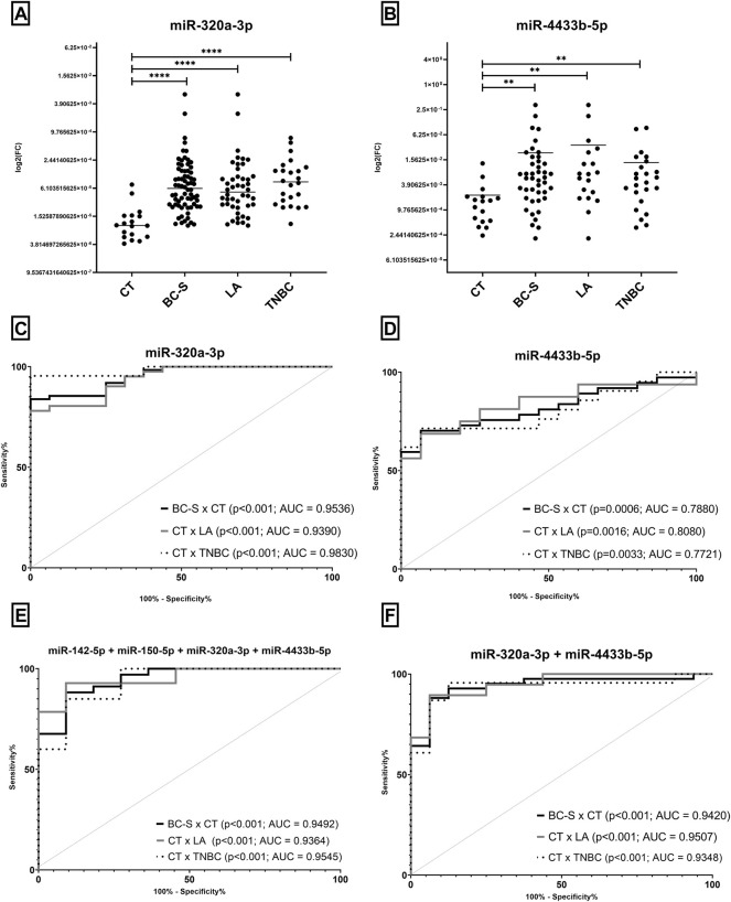
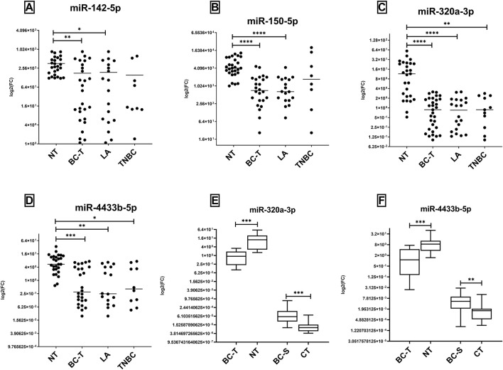
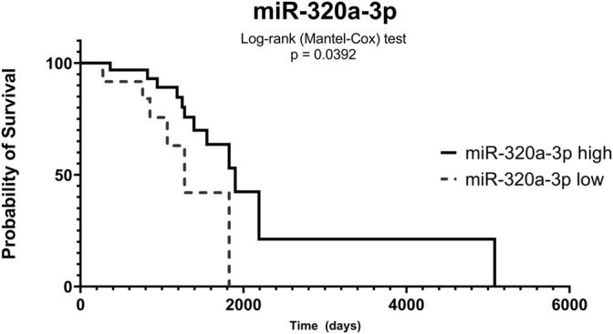
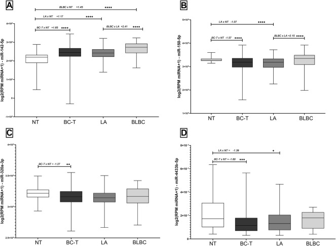
Breast cancer (BC) is a heterogeneous disease, and establishing biomarkers is essential to patient management. We previously described that extracellular vesicle-derived miRNAs (EV-miRNAs) miR-142-5p, miR-150-5p, miR-320a, and miR-4433b-5p in serum discriminated BC from control samples, either alone or combined in a panel. Using these previously described markers, we intend to evaluate whether the same markers identified in EVs are also potential biomarkers in tissue and serum. Expression analysis using RT-qPCR was performed using serum of 67 breast cancer patients (BC-S), 19 serum controls (CT), 83 fresh tumor tissues (BC-T), and 29 adjacent nontumor tissue samples (NT). In addition, analysis from The Cancer Genome Atlas (TCGA) data (832 BC-T and 136 NT) was performed. In all comparisons, we found concordant high expression levels of miR-320a and miR-4433b-5p in BC-S compared to CT in both EVs and cell-free miRNAs (cf-miRNAs). Although miR-150-5p and miR-142-5p were not found to be differentially expressed in serum, panels including these miRNAs improved sensitivity and specificity, supporting our previous findings in EVs. Fresh tissue and data from the TCGA database had, in most comparisons, an opposite behavior when compared to serum and EVs: lower levels of all miRNAs in BC-T than those in NT samples. TCGA analyses revealed reduced expression levels of miR-150-5p and miR-320a-3p in BC-T than those in NT samples and the overexpression of miR-142-5p in BC-T, unlike our RT-qPCR results from tissue in the Brazilian cohort. The fresh tissue analysis showed that all miRNAs individually could discriminate between BC-T and NT in the Brazilian cohort, with high sensitivity and sensibility. Furthermore, combining panels showed higher AUC values and improved sensitivity and specificity. In addition, lower levels of miR-320a-3p in serum were associated with poor overall survival in BC Brazilian patients. In summary, we observed that miR-320a and miR-4433b-5p distinguished BC from controls with high specificity and sensibility, regardless of the sample source. In addition, lower levels of miR-150-5p and higher levels of miR-142-5p were statistically significant biomarkers in tissue, according to TCGA. When combined in panels, all combinations could distinguish BC patients from controls. These results highlight a potential application of these miRNAs as BC biomarkers.
Keywords: biomarkers; breast cancer; miR-142-5p; miR-150-5p; miR-320a; miR-4433b-5p.
Copyright © 2022 Carvalho, Brasil, Jucoski, Adamoski, Lima, Spautz, Anselmi, Ozawa, Cavalli, Carvalho de Oliveira, Gradia and Ribeiro.
Conflict of interest statement
The authors declare that the research was conducted in the absence of any commercial or financial relationships that could be construed as a potential conflict of interest.
Figures
Similar articles
-
Identification of miRNAs Enriched in Extracellular Vesicles Derived from Serum Samples of Breast Cancer Patients.Biomolecules. 2020 Jan 16;10(1):150. doi: 10.3390/biom10010150. Biomolecules. 2020. PMID: 31963351 Free PMC article.
-
Circulating microRNAs from the miR-106a-363 cluster on chromosome X as novel diagnostic biomarkers for breast cancer.Breast Cancer Res Treat. 2018 Jul;170(2):257-270. doi: 10.1007/s10549-018-4757-3. Epub 2018 Mar 20. Breast Cancer Res Treat. 2018. PMID: 29557526 Free PMC article.
-
Urinary Exosomal miRNAs as biomarkers of bladder Cancer and experimental verification of mechanism of miR-93-5p in bladder Cancer.BMC Cancer. 2021 Dec 3;21(1):1293. doi: 10.1186/s12885-021-08926-x. BMC Cancer. 2021. PMID: 34861847 Free PMC article.
-
Identification of differentially expressed circulating serum microRNA for the diagnosis and prognosis of Indian non-small cell lung cancer patients.Curr Probl Cancer. 2020 Aug;44(4):100540. doi: 10.1016/j.currproblcancer.2020.100540. Epub 2020 Jan 23. Curr Probl Cancer. 2020. PMID: 32007320 Review.
-
MicroRNA Expression in Endometrial Cancer: Current Knowledge and Therapeutic Implications.Medicina (Kaunas). 2024 Mar 15;60(3):486. doi: 10.3390/medicina60030486. Medicina (Kaunas). 2024. PMID: 38541212 Free PMC article.
Cited by
-
Circulating Small EVs miRNAs as Predictors of Pathological Response to Neo-Adjuvant Therapy in Breast Cancer Patients.Int J Mol Sci. 2022 Oct 20;23(20):12625. doi: 10.3390/ijms232012625. Int J Mol Sci. 2022. PMID: 36293478 Free PMC article.
-
Diagnostic and prognostic significance of miR-320a-3p in patients with chronic heart failure.BMC Cardiovasc Disord. 2024 Jun 17;24(1):308. doi: 10.1186/s12872-024-03966-0. BMC Cardiovasc Disord. 2024. PMID: 38886631 Free PMC article.
-
LncRNA OIP5-AS1 Upregulates the Cyclin D2 Levels to Promote Metastasis of Breast Cancer by Targeting miR-150-5p.Appl Biochem Biotechnol. 2024 Dec;196(12):8627-8644. doi: 10.1007/s12010-024-04992-6. Epub 2024 Jun 18. Appl Biochem Biotechnol. 2024. PMID: 38888699
-
Tumor-derived small extracellular vesicles facilitate omental metastasis of ovarian cancer by triggering activation of mesenchymal stem cells.Cell Commun Signal. 2024 Jan 17;22(1):47. doi: 10.1186/s12964-023-01413-9. Cell Commun Signal. 2024. PMID: 38233863 Free PMC article.
-
Combinational treatments of RNA interference and extracellular vesicles in the spinocerebellar ataxia.Front Mol Neurosci. 2022 Oct 13;15:1043947. doi: 10.3389/fnmol.2022.1043947. eCollection 2022. Front Mol Neurosci. 2022. PMID: 36311034 Free PMC article. Review.
References
LinkOut - more resources
Full Text Sources
Research Materials