

Granulosa Cells Improved Mare Oocyte Cytoplasmic Maturation by Providing Collagens
- PMID: 35846364
- PMCID: PMC9280134
- DOI: 10.3389/fcell.2022.914735
Granulosa Cells Improved Mare Oocyte Cytoplasmic Maturation by Providing Collagens
Abstract
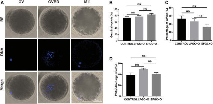
Assisted reproductive technology has important clinical applications and commercial values in the horse industry. However, this approach is limited largely by the low efficiency of oocyte in vitro maturation (IVM), especially cytoplasmic maturation. To improve the efficiency of mare oocyte IVM, we evaluated the effects of co-culture with cumulus-oocyte complexes (COCs) and granulosa cells (GCs) from follicles with small (<15 mm) and large diameters (>35 mm). Our results showed that oocyte nucleus maturation was not significantly improved by co-culturing with GCs. Interestingly, the cytoplasmic maturation of oocytes, defined by the distribution of cortical granules and mitochondria, as well as reactive oxygen species (ROS) levels, improved dramatically by co-culture with GCs, especially those derived from small follicles. Moreover, GCs promoted cumulus cell expansion by upregulating the expression of BMP15 in oocytes. To determine the mechanism underlying the effects of GCs, the transcriptomes of GCs from large and small follicles were compared. Expression levels of COL1A2, COL6A1, and COL6A2 were significantly higher in GCs from small follicles than in those from large follicles. These three genes were enriched in the extracellular matrix proteins-receptor interaction pathway and were involved in the regulation of collagens. Taken together, our results suggest that co-culture with GCs is beneficial to oocyte cytoplasmic maturation, and the increased expression of COL1A2, COL6A1, and COL6A2 improve the mare oocyte IVM system via the regulation of collagen.
Keywords: BMP15; collagens; cytoplasm maturation; granulosa cells; mare; oocyte.
Copyright © 2022 Zhu, Zhao, Xu, Zhang, Zhu, Pan and Huang.
Conflict of interest statement
The authors declare that the research was conducted in the absence of any commercial or financial relationships that could be construed as a potential conflict of interest.
Figures


Similar articles
-
An improved IVM method for cumulus-oocyte complexes from small follicles in polycystic ovary syndrome patients enhances oocyte competence and embryo yield.Hum Reprod. 2017 Oct 1;32(10):2056-2068. doi: 10.1093/humrep/dex262. Hum Reprod. 2017. PMID: 28938744
-
MicroRNAs transfected into granulosa cells may regulate oocyte meiotic competence during in vitro maturation of mouse follicles.Hum Reprod. 2013 Nov;28(11):3050-61. doi: 10.1093/humrep/det338. Epub 2013 Aug 26. Hum Reprod. 2013. PMID: 23980055
-
In vitro maturation on ovarian granulosa cells encapsulated in agarose matrix improves developmental competence of porcine oocytes.Theriogenology. 2021 Apr 1;164:42-50. doi: 10.1016/j.theriogenology.2021.01.008. Epub 2021 Jan 21. Theriogenology. 2021. PMID: 33540369
-
Despite the donor's age, human adipose-derived stem cells enhance the maturation and development rates of porcine oocytes in a co-culture system.Theriogenology. 2018 Jul 15;115:57-64. doi: 10.1016/j.theriogenology.2017.12.024. Epub 2017 Dec 12. Theriogenology. 2018. PMID: 29709724 Review.
-
Molecular characteristics of oocytes and somatic cells of follicles at different sizes that influence in vitro oocyte maturation and embryo production.Domest Anim Endocrinol. 2021 Jan;74:106485. doi: 10.1016/j.domaniend.2020.106485. Epub 2020 Apr 21. Domest Anim Endocrinol. 2021. PMID: 32858464 Review.
Cited by
-
Unravelling the Signature Follicular Fluid Metabolites in Dairy Cattle Follicles Growing Under Negative Energy Balance: An In Vitro Approach.Int J Mol Sci. 2024 Nov 25;25(23):12629. doi: 10.3390/ijms252312629. Int J Mol Sci. 2024. PMID: 39684341 Free PMC article.
-
3D hUC-MSC spheroids exhibit superior resistance to autophagy and apoptosis of granulosa cells in POF rat model.Reproduction. 2024 Jul 13;168(2):e230496. doi: 10.1530/REP-23-0496. Print 2024 Aug 1. Reproduction. 2024. PMID: 38912966 Free PMC article.
-
Importance of Antioxidant Supplementation during In Vitro Maturation of Mammalian Oocytes.Vet Sci. 2022 Aug 18;9(8):439. doi: 10.3390/vetsci9080439. Vet Sci. 2022. PMID: 36006354 Free PMC article. Review.
References
-
- Adeldust H., Zeinoaldini S., Kohram H., Amiri Roudbar M., Daliri Joupari M. (2015). In Vitro maturation of Ovine Oocyte in a Modified Granulosa Cells Co-culture System and Alpha-Tocopherol Supplementation: Effects on Nuclear Maturation and Cleavage. J. Anim. Sci. Technol. 57, 27. 10.1186/s40781-015-0061-5 - DOI - PMC - PubMed
-
- Caixeta E. S., Sutton-Mcdowall M. L., Gilchrist R. B., Thompson J. G., Price C. A., Machado M. F., et al. (2013). Bone Morphogenetic Protein 15 and Fibroblast Growth Factor 10 Enhance Cumulus Expansion, Glucose Uptake, and Expression of Genes in the Ovulatory Cascade during In Vitro Maturation of Bovine Cumulus-Oocyte Complexes. Reproduction 146, 27–35. 10.1530/rep-13-0079 - DOI - PubMed
LinkOut - more resources
Full Text Sources
Miscellaneous