

Transorbital hybrid approach for endovascular occlusion of indirect carotid-cavernous fistulas-Case report and systematic literature review
- PMID: 35846510
- PMCID: PMC9283805
- DOI: 10.1016/j.radcr.2022.06.043
Transorbital hybrid approach for endovascular occlusion of indirect carotid-cavernous fistulas-Case report and systematic literature review
Abstract
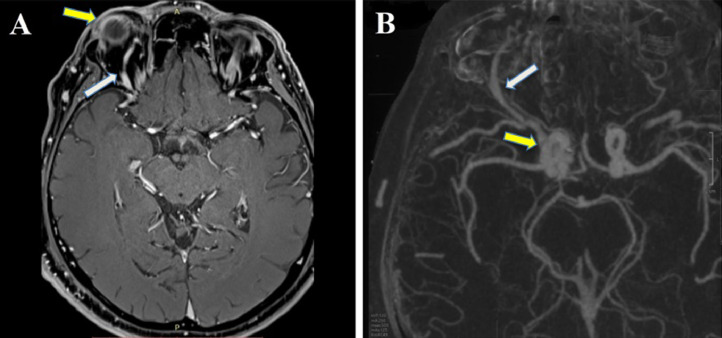
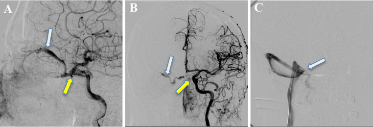
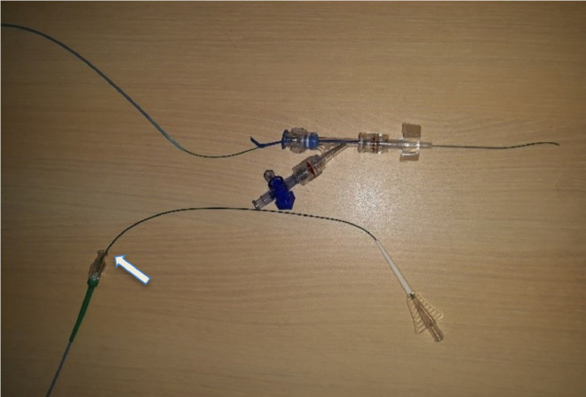
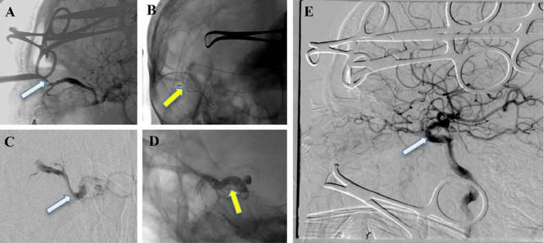
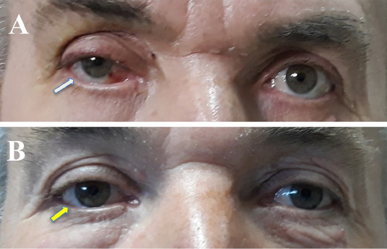
Carotid-cavernous fistulas (CCF) are vascular malformations characterized by an aberrant shunt between one or more sources of arterial inflow and the cavernous sinus (CS). They are subdivided into direct and indirect fistulas. This last one, called dural CCF involve dural fistulous connections between branches of the internal carotid artery or the external carotid artery. When conventional routes are not eligible, surgical exposure of the vein is the only access to the fistula. We present the case of a patient successfully treated for right sided dural CCF, by a hybrid approach. Furthermore, through a literature review, we analyze the possible risks and benefits associated with this approach.
Keywords: Dural carotid-cavernous fistula; Embolization; Hybrid approach; Superior ophthalmic vein cannulation.
© 2022 The Authors. Published by Elsevier Inc. on behalf of University of Washington.
Figures
Similar articles
-
Surgical Sparing and Pairing Endovascular Interventions for Carotid-Cavernous Fistula: Case Series and Review of the Literature.World Neurosurg. 2020 Aug;140:18-25. doi: 10.1016/j.wneu.2020.05.013. Epub 2020 May 11. World Neurosurg. 2020. PMID: 32437988 Review.
-
Transorbital Approach for Endovascular Occlusion of Carotid-Cavernous Fistulas: Technical Note and Review of the Literature.Cureus. 2017 Jan 12;9(1):e976. doi: 10.7759/cureus.976. Cureus. 2017. PMID: 28191380 Free PMC article.
-
Cavernous sinus dural fistulae treated by transvenous approach through the facial vein: report of seven cases and review of the literature.AJNR Am J Neuroradiol. 2003 Jun-Jul;24(6):1240-6. AJNR Am J Neuroradiol. 2003. PMID: 12812963 Free PMC article.
-
Endoscope-assisted transsphenoidal puncture of the cavernous sinus for embolization of carotid-cavernous fistula in a neurosurgical hybrid operating suite.J Neurosurg. 2017 Aug;127(2):327-331. doi: 10.3171/2016.5.JNS16493. Epub 2016 Aug 5. J Neurosurg. 2017. PMID: 27494822
-
Endovascular approaches for the treatment of dural carotid-cavernous fistulas: A systematic review.Interv Neuroradiol. 2024 Aug 8:15910199241272595. doi: 10.1177/15910199241272595. Online ahead of print. Interv Neuroradiol. 2024. PMID: 39113637 Free PMC article. Review.
Cited by
-
Editorial: Hybrid (combined endovascular and microsurgical) treatments for cerebrovascular diseases.Front Neurol. 2024 Mar 11;15:1378269. doi: 10.3389/fneur.2024.1378269. eCollection 2024. Front Neurol. 2024. PMID: 38533415 Free PMC article. No abstract available.
References
-
- Leone G., Renieri L., Enriquez-Marulanda A., Dmytriw A.A., Nappini S., Laiso A., et al. Carotid Cavernous Fistulas and Dural Arteriovenous Fistulas of the Cavernous Sinus: Validation of a New Classification According to Venous Drainage. World Neurosurg. Aug 2019;128:e621–e631. doi: 10.1016/j.wneu.2019.04.220. - DOI - PubMed
Publication types
LinkOut - more resources
Full Text Sources
