

Imaging of Group B Streptococcus infection in pyonephrosis: a case report
- PMID: 35846511
- PMCID: PMC9284314
- DOI: 10.1016/j.radcr.2022.06.014
Imaging of Group B Streptococcus infection in pyonephrosis: a case report
Abstract
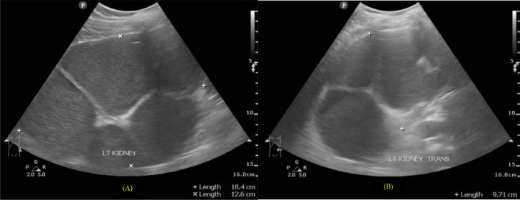
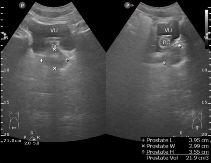
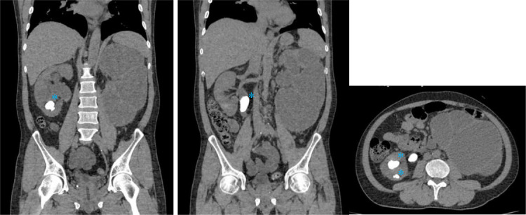
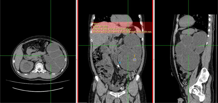
Introduction: Pyonephrosis is hydronephrosis accompanied by a bacterial infection in the kidney, causing suppurative destruction of the renal parenchyma; this condition is an emergency and usually associated with stones or chronic urinary tract infections. Urinalysis is typically inaccurate for establishing the diagnosis, as bacteriuria may not manifest due to ureteral obstruction. Case report: We reported a 55-year-old male patient with flanks pain and an account of stone expulsion. Based on history taking, physical examination, radiology examinations, and percutaneous nephrotomy, we concluded a diagnosis of pyonephrosis causing by Streptococcus agalactiae as known as Group B Streptococcus. Discussion: While both US and CT scan guided the early diagnosis, CT was more accurate as it is able to capture the renal function and the underlying cause of obstruction. Pyonephrosis was described as having a pus collection in the pelvicalyceal system, cortex thinning, and the appearance of stones. Conclusion: Pyonephrosis is a rare emergency, and many clinicians find it challenging to recognize since the presentations are frequently nonspecific. In order to prevent renal failure and the spread of bacteremia that entails life-threatening urosepsis, acquiring imaging knowledge (sonography and CT) and other findings are indispensable in determining this entity.
Keywords: GBS; Pyonephrosis; Streptococcus agalactiae, Imaging.
© 2022 The Authors.
Figures


Similar articles
-
A Nautilus kidney.Clin Exp Nephrol. 2020 Oct;24(10):971-972. doi: 10.1007/s10157-020-01915-7. Epub 2020 Jun 26. Clin Exp Nephrol. 2020. PMID: 32592079
-
Pyonephrosis as the first symptom of congenital hydronephrosis in a 6-year old girl.Pol Merkur Lekarski. 2018 Apr 23;44(262):196-200. Pol Merkur Lekarski. 2018. PMID: 29775448
-
Giant Pyonephrosis Due to Ureteropelvic Junction Obstruction: A Case Report.J Clin Diagn Res. 2017 Aug;11(8):PD17-PD18. doi: 10.7860/JCDR/2017/26103.10506. Epub 2017 Aug 1. J Clin Diagn Res. 2017. PMID: 28969204 Free PMC article.
-
Case report of ureter obturator hernia and literature review analysis.BMC Urol. 2021 May 29;21(1):86. doi: 10.1186/s12894-021-00851-2. BMC Urol. 2021. PMID: 34051776 Free PMC article. Review.
-
Pyonephrosis Ultrasound and Computed Tomography Features: A Pictorial Review.Diagnostics (Basel). 2021 Feb 17;11(2):331. doi: 10.3390/diagnostics11020331. Diagnostics (Basel). 2021. PMID: 33671431 Free PMC article. Review.
Cited by
-
Missing Renal Stone Diagnosis in Dementia Patients With Recurrent Urinary Tract Infections: A Case Report and Literature Review.Cureus. 2024 Apr 24;16(4):e58908. doi: 10.7759/cureus.58908. eCollection 2024 Apr. Cureus. 2024. PMID: 38800185 Free PMC article.
-
Peribacillus simplex and Klebsiella pneumoniae responsible for pyonephrosis with secondary psoas abscess: a case report.Access Microbiol. 2025 Jan 14;7(1):000911.v3. doi: 10.1099/acmi.0.000911.v3. eCollection 2025. Access Microbiol. 2025. PMID: 39810798 Free PMC article.
References
-
- Tublin M, Levine D, Thurston W, Wilson SR. In: Diagnostic Ultrasound. 5th ed. Rumack CM, Levine D, editors. Elsevier; Philadelpia, PA: 2018. The kidney and urinary tract; p. 325.
-
- Farley MM, Harvey RC, Stull T, Smith JD, et al. A population-based assessment of invasive disease due to Group B Streptococcus in nonpregnant adults. N Engl J Med. 1993;328(25):1807–1811. - PubMed
-
- Cooper KL, Badalato GM, Rutman MP. Campbell-Walsh Urology. Elsevier; Philadelpia, PA: 2021. Infections of the urinary tract; p. 1174. Partin AW, editor.
Publication types
LinkOut - more resources
Full Text Sources
