. 2022 Jun 30:16:912030.
doi: 10.3389/fncel.2022.912030. eCollection 2022.

Neuronal Adenosine A1 Receptor is Critical for Olfactory Function but Unable to Attenuate Olfactory Dysfunction in Neuroinflammation

Affiliations

Neuronal Adenosine A1 Receptor is Critical for Olfactory Function but Unable to Attenuate Olfactory Dysfunction in Neuroinflammation

Charlotte Schubert et al. Front Cell Neurosci. .

Abstract

Adenine nucleotides, such as adenosine triphosphate (ATP), adenosine diphosphate (ADP), as well as the nucleoside adenosine are important modulators of neuronal function by engaging P1 and P2 purinergic receptors. In mitral cells, signaling of the G protein-coupled P1 receptor adenosine 1 receptor (A1R) affects the olfactory sensory pathway by regulating high voltage-activated calcium channels and two-pore domain potassium (K2P) channels. The inflammation of the central nervous system (CNS) impairs the olfactory function and gives rise to large amounts of extracellular ATP and adenosine, which act as pro-inflammatory and anti-inflammatory mediators, respectively. However, it is unclear whether neuronal A1R in the olfactory bulb modulates the sensory function and how this is impacted by inflammation. Here, we show that signaling via neuronal A1R is important for the physiological olfactory function, while it cannot counteract inflammation-induced hyperexcitability and olfactory deficit. Using neuron-specific A1R-deficient mice in patch-clamp recordings, we found that adenosine modulates spontaneous dendro-dendritic signaling in mitral and granule cells via A1R. Furthermore, neuronal A1R deficiency resulted in olfactory dysfunction in two separate olfactory tests. In mice with experimental autoimmune encephalomyelitis (EAE), we detected immune cell infiltration and microglia activation in the olfactory bulb as well as hyperexcitability of mitral cells and olfactory dysfunction. However, neuron-specific A1R activity was unable to attenuate glutamate excitotoxicity in the primary olfactory bulb neurons in vitro or EAE-induced olfactory dysfunction and disease severity in vivo. Together, we demonstrate that A1R modulates the dendro-dendritic inhibition (DDI) at the site of mitral and granule cells and impacts the processing of the olfactory sensory information, while A1R activity was unable to counteract inflammation-induced hyperexcitability.

Keywords: A1R; EAE; adenosine; mitral cells; neuroprotection; olfactory bulb; olfactory dysfunction; purinergic signaling.


Conflict of interest statement

The authors declare that the research was conducted in the absence of any commercial or financial relationships that could be construed as a potential conflict of interest.

Figures

FIGURE 1
FIGURE 1
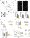
A1Rflx/flx;SNAP25Cre results in neuron-specific A1R knockout. (A) HA-tagged reporter expression in neuron-specific A1R knockout mouse line A1Rflx/flx;Tag;SNAP25Cre indicates wide distribution of Cre expression in the main olfactory bulb. Representative image shows a sagittal view of the main olfactory bulb comprising the granular cell layer (GCL), internal plexiform layer (not marked), mitral cell layer (MCL), external plexiform layer (EPL), and the glomerular layer (GL) as indicated by different colors. HA-tag was stained with anti-HA antibody. (B) Quantitative qPCR of Adora1 (coding for the A1R) mRNA expression of olfactory bulbs and spleens (BF10 = 0.538) isolated from A1Rflx/flx (n = 3) and A1Rflx/flx;SNAP25Cre (n = 3). (C) Quantitative qPCR mRNA expression of P1 receptor genes Adora1, Adora2a (BF10 = 0.646), Adora2b (BF10 = 0.624), and Adora3 (BF10 = 0.565) in the olfactory bulb of A1Rflx/flx (n = 3) and A1Rflx/flx;SNAP25Cre (n = 3) mice. (D) Mitral cells analyzed by marker Reelin in the mitral cell layer in A1Rflx/flx (n = 5) and A1Rflx/flx;SNAP25Cre (n = 5) mice (BF10 = 0.337). (E) Outward shift of the holding current in the mitral cell patch clamp recordings evoked by the application of 100 μM of adenosine (ADO) in A1Rflx/flx (n = 7 slices from 5 mice) and A1Rflx/flx;SNAP25Cre (n = 5 slices from 4 mice) (MWU; U = 0, P = 0.0025). Representative traces in gray (A1Rflx/flx) and olive (A1Rflx/flx;SNAP25Cre). (F) Input resistance was measured in patch clamp recordings of mitral cells from A1Rflx/flx (n = 7 slices from 5 mice) and A1Rflx/flxSNAP25Cre (n = 5 slices from 4 mice) mice, BF10 = 0.486. (G) Progression of the F/I plot shows excitability of mitral cells from A1Rflx/flx (n = 7 slices from 5 mice) mice vs. A1Rflx/flx;SNAP25Cre (n = 5 slices from 4 mice) (BF10 = 0.480-0.537). Data points display mean ± s.e.m. **P < 0.01, ***P < 0.001.
FIGURE 2
FIGURE 2
Adenosine modulates microcircuits in the external plexiform layer of the main olfactory bulb via A1R. (A) Schematic presentation of main olfactory bulb with principal neurons (MC, TC) and local interneurons [periglomerular neurons (PG), short axon cells (sAC), parvalbumin neurons (PV), and granule cells (GC)]. Inset: Mechanism of dendro-dendritic signaling at the reciprocal MC-GC synapse. (B) Representative images of synaptic density in the external plexiform layer (EPL) of the main olfactory bulb in A1R-proficient (A1Rflx/flx) and A1R-deficient (A1Rflx/flx;SNAP25Cre) mice. Immunohistochemical stainings comprised the presynaptic protein synapsin1 and the postsynaptic protein PSD95. (C) Immunohistochemical analysis of synapsin1, PSD95 (BF10 = 0.518), and co-expression of synapsin1 and PSD95 in the EPL in A1Rflx/flx and A1Rflx/flx;SNAP25Cre (n = 6 per group). (D) Whole cell current recordings of granule cells. Number of synaptic inputs quantified as the sum of sPSC events per minute in A1Rflx/flx (n = 18) compared to A1Rflx/flx;SNAP25Cre (n = 9). MWU; U = 40, P = 0.035. (E) Whole cell current recordings of mitral cells. Number of synaptic inputs quantified as the sum of sPSC events per minute (A1Rflx/flx n = 9; A1Rflx/flx;SNAP25Cre n = 8), BF10 = 0.476. (F) Frequency of sPSCs in mitral cells and granule cells after the application of 100 μM of adenosine (mean of second 130–170 of recording) compared to baseline (mean of second 30–70 of recording) in A1Rflx/flx (n = 10 in granule cells, n = 9 in mitral cells) and A1Rflx/flx;SNAP25Cre (n = 10 in granule cells, n = 9 in mitral cells). Adenosine-dependent effect on sPSC frequency was analyzed by the Wilcoxon ranking test. For A1R-proficient cells, P = 0.00195 (granule cells) and P = 0.00391 (mitral cells); for A1R-deficient cells, BF10 (paired test) = 0.322 (granule cells) and BF10 (paired test) = 0.793 (mitral cells). Genotype comparison was done by Mann–Whitney U test. Adenosine effect A1Rflx/flx vs. A1Rflx/flx;SNAP25Cre for granule cells (U = 6, P = 0.003) and mitral cells (U = 15, P = 0.024). (G) Frequency of sPSCs in mitral cells normalized to baseline over the time course of >7 min. Representative traces showing sPSC of mitral cells under baseline condition and after treatment with 100 μM of adenosine at timespoints indicated by arrows. *P < 0.05, ***P < 0.001.
FIGURE 3
FIGURE 3
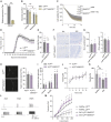
A1R deficiency results in olfactory dysfunction. (A) Graphical workflow of olfactory detection test. A1Rflx/flx and A1Rflx/flx;SNAP25Cre were exposed to empty tubes (black), or tubes perfumed with a concentrate of vanilla (green) or almond (orange). Each trial lasted 4 min. (B) Quantification of olfactory detection tests. Preference to new odor analyzed by the time of sniffing at the respective side (s) (n = 10 per group, BF10 = 0.361). (C) Events of rearing during each trial in olfactory detection test of A1Rflx/flx and A1Rflx/flx;SNAP25Cre mice (BF10 = 0.439–0.473). (D) TMT-based olfactory test. Time of freezing in percent in bins of 2 min in A1Rflx/flx (n = 11) and A1Rflx/flx;SNAP25Cre (n = 6). Bonferroni post-hoc test after a mixed multifactorial ANOVA; *P < 0.05.
FIGURE 4
FIGURE 4
Neuroinflammation results in olfactory dysfunction and hyperexcitability that is not influenced by neuronal A1R. (A) Adora1 expression in primary neuronal cultures of olfactory bulb neurons DIV 14–16 of A1Rflx/flx (gray) and A1Rflx/flx;SNAP25Cre (beige) (n = 3 per group). (B) Neuronal survival in vitro measured by MAP2-positive cells after treatment with 5 μM of glutamate for 6 h in A1Rflx/flx and A1Rflx/flx;SNAP25Cre (n = 3 biological replicates with 2–3 technical replicates). (C) Luminescence-based measurement of cell viability over 15 h treated with 10 μM of adenosine (ADO) and 5 μM of glutamate or medium as control (n = 4 biological replicates per group with at least 5 technical replicates each). (D) Clinical time course of experimental autoimmune encephalomyelitis (EAE) over 30 days (n = 18 per group) in A1R-proficient (A1Rflx/flx) and A1R-deficient (A1Rflx/flx;SNAP25Cre) mice. (E) Disease severity in EAE analyzed by area under the curve (BF10 = 0.384). (F) Representative images and (G) quantification of microglia activation measured by Iba1 (BF10 = 0.506), and T-cell infiltration evaluated by CD3-positive cells/mm2 (BF10 = 0.476) in the main olfactory bulb in coronal slices (n = 5 per group). (H) Reelin-positive mitral cells per mm mitral cell layer at day 30 post-immunization (p.i.) (n = 5 per group, BF10 = 0.673). (I) Olfactory detection test at the early phase of EAE (day 10–12 p.i.) in A1Rflx/flx n = 6 and A1Rflx/flxSNAP25Cre n = 7. As mice were exposed to odors at two sides of the cage (first trial empty control, second trial vanilla on the right side, third trial almond on the left side where vanilla was reintroduced on the right side), preference to one side of the cage has been measured by means of the ratio between the sides. (J) TMT-based olfactory testing measured by the percentage of freezing in bins of 2 min in EAE (A1Rflx/flx n = 10 and A1Rflx/flx;SNAP25Cre n = 8). (K) TMT-based olfactory behavioral test in healthy animals and at the early phase of EAE. Percentage of freezing after 10 min in A1Rflx/flx (healthy n = 11 in light gray, EAE n = 10 in gray) and A1Rflx/flx;SNAP25Cre mice (healthy n = 6 in light lavender, EAE n = 8 in lavender). (L) Representative traces of mitral cell action potentials in healthy control (light gray) and acute phase of EAE (dark gray). (M) Increase of frequency of action potentials of mitral cells depending on the injected current in healthy (n = 7) and acute EAE (n = 5) slice preparations of A1Rflx/flx (healthy n = 7; EAE n = 5) and A1Rflx/flx;SNAP25Cre (healthy n = 5; EAE n = 7) mice, analyzed by Mann–Whitney U-test. Statistical analysis was performed by two-way ANOVA in repetitive measurements (C,D,J,L,M) and unpaired t-test, if not stated otherwise; *P < 0.05, **P < 0.01.

Similar articles

Cited by

References

    1. Abraham N. M., Egger V., Shimshek D. R., Renden R., Fukunaga I., Sprengel R., et al. (2010). Synaptic inhibition in the olfactory bulb accelerates odor discrimination in mice. Neuron 65 399–411. 10.1016/J.NEURON.2010.01.009 - DOI - PMC - PubMed
    1. Attfield K. E., Jensen L. T., Kaufmann M., Friese M. A., Fugger L. (2022). The immunology of multiple sclerosis. Nat. Rev. Immunol. 28 29–45. 10.1038/S41577-022-00718-Z - DOI - PubMed
    1. Ballesteros-Yáñez I., Castillo C. A., Merighi S., Gessi S. (2018). The role of adenosine receptors in psychostimulant addiction. Front. Pharmacol. 8:985. 10.3389/fphar.2017.00985 - DOI - PMC - PubMed
    1. Balu R., Pressler R. T., Strowbridge B. W. (2007). Multiple modes of synaptic excitation of olfactory bulb granule cells. J. Neurosci. 27:5621. 10.1523/JNEUROSCI.4630-06.2007 - DOI - PMC - PubMed
    1. Banie A. P., Nicholls D. G. (1993). Adenosine A1 receptor inhibition of glutamate exocytosis and protein kinase c-mediated decoupling. J. Neurochem. 60 1081–1086. 10.1111/j.1471-4159.1993.tb03257.x - DOI - PubMed

LinkOut - more resources