

The Shape of μ-How Morphological Analyses Shape the Study of Microglia
- PMID: 35846562
- PMCID: PMC9276927
- DOI: 10.3389/fncel.2022.942462
The Shape of μ-How Morphological Analyses Shape the Study of Microglia
Abstract
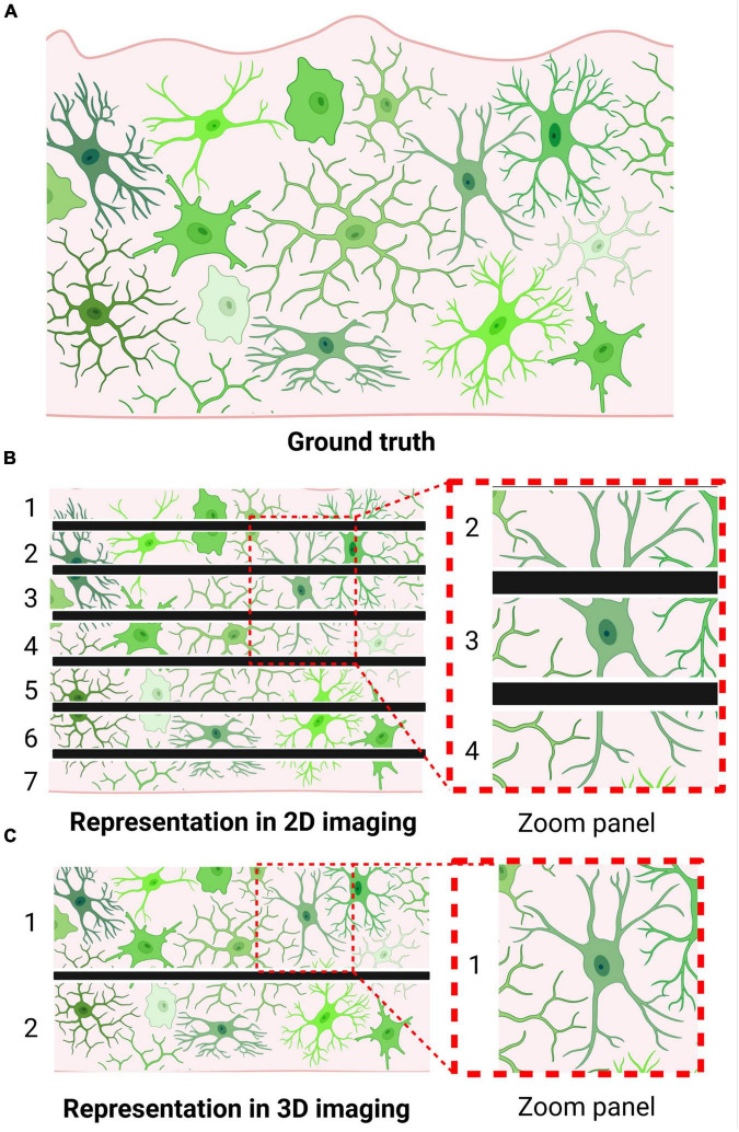
Microglia, the innate immune cells of the CNS parenchyma, serve as the first line of defense in a myriad of neurodevelopmental, neurodegenerative, and neuroinflammatory conditions. In response to the peripheral inflammation, circulating mediators, and other external signals that are produced by these conditions, microglia dynamically employ different transcriptional programs as well as morphological adaptations to maintain homeostasis. To understand these cells' function, the field has established a number of essential analysis approaches, such as gene expression, cell quantification, and morphological reconstruction. Although high-throughput approaches are becoming commonplace in regard to other types of analyses (e.g., single-cell scRNA-seq), a similar standard for morphological reconstruction has yet to be established. In this review, we offer an overview of microglial morphological analysis methods, exploring the advantages and disadvantages of each, highlighting a number of key studies, and emphasizing how morphological analysis has significantly contributed to our understanding of microglial function in the CNS parenchyma. In doing so, we advocate for the use of unbiased, automated morphological reconstruction approaches in future studies, in order to capitalize on the valuable information embedded in the cellular structures microglia inhabit.
Keywords: 3D reconstruction; automated image analysis; electron microscopy; in vivo imaging; microglia; microglia morphology.
Copyright © 2022 Bosch and Kierdorf.
Conflict of interest statement
The authors declare that the research was conducted in the absence of any commercial or financial relationships that could be construed as a potential conflict of interest.
Figures
References
-
- Achúcarro N. (1909). Cellules Allongées Et Stäbechenzellen: Cellules Neurogliques Et Cellules Granulo-Adipeuses Á La Corne D’ammon Du Lapin. Paris: Nicolás Moya.
-
- Bloomfield P. S., Bonsall D., Wells L., Dormann D., Howes O., De Paola V. (2018). The effects of haloperidol on microglial morphology and translocator protein levels: an in vivo study in rats using an automated cell evaluation pipeline. J. Psychopharmacol. 32 1264–1272. 10.1177/0269881118788830 - DOI - PubMed
Publication types
LinkOut - more resources
Full Text Sources
Research Materials