. 2022 Jun 30:16:913009.
doi: 10.3389/fncel.2022.913009. eCollection 2022.

Exosomes Derived From Umbilical Cord Mesenchymal Stem Cells Treat Cutaneous Nerve Damage and Promote Wound Healing

Affiliations

Exosomes Derived From Umbilical Cord Mesenchymal Stem Cells Treat Cutaneous Nerve Damage and Promote Wound Healing

Ziying Zhu et al. Front Cell Neurosci. .

Abstract

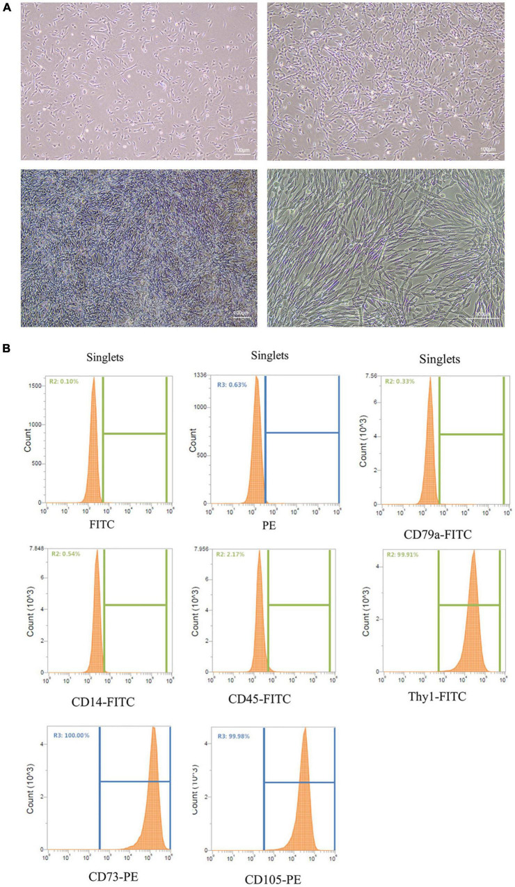
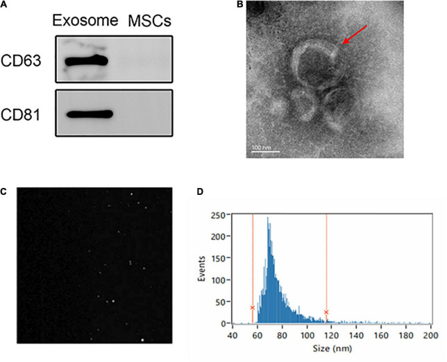
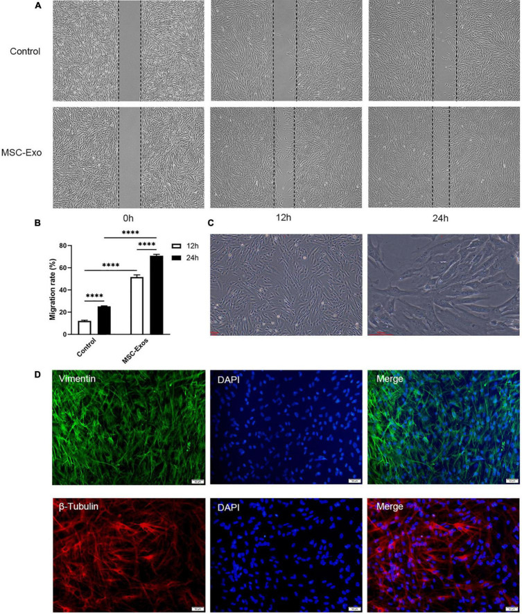
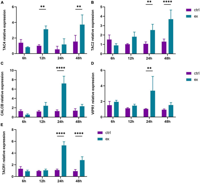
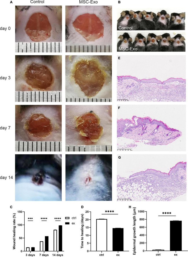
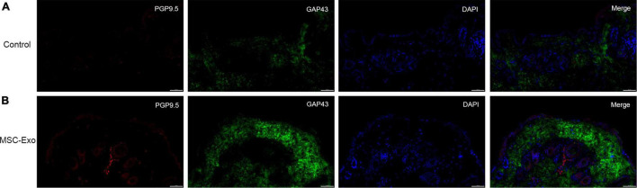
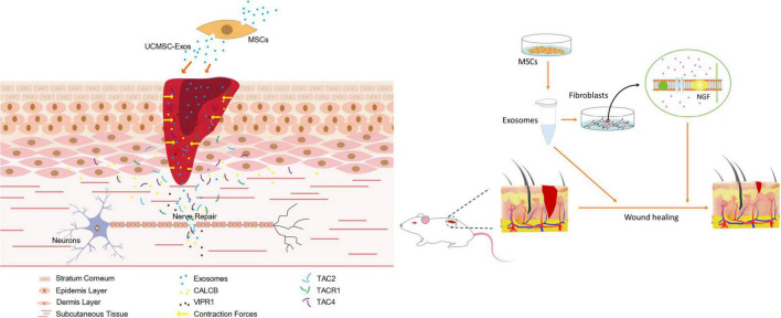
Wound repair is a key step in the treatment of skin injury caused by burn, surgery, and trauma. Various stem cells have been proven to promote wound healing and skin regeneration as candidate seed cells. Therefore, exosomes derived from stem cells are emerging as a promising method for wound repair. However, the mechanism by which exosomes promote wound repair is still unclear. In this study, we reported that exosomes derived from umbilical cord mesenchymal stem cells (UC-MSCs) promote wound healing and skin regeneration by treating cutaneous nerve damage. The results revealed that UC-MSCs exosomes (UC-MSC-Exo) promote the growth and migration of dermal fibroblast cells. In in vitro culture, dermal fibroblasts could promote to nerve cells and secrete nerve growth factors when stimulated by exosomes. During the repair process UC-MSC-Exo accelerated the recruitment of fibroblasts at the site of trauma and significantly enhanced cutaneous nerve regeneration in vivo. Interestingly, it was found that UC-MSC-Exo could promote wound healing and skin regeneration by recruiting fibroblasts, stimulating them to secrete nerve growth factors (NGFs) and promoting skin nerve regeneration. Therefore, we concluded that UC-MSC-Exo promote cutaneous nerve repair, which may play an important role in wound repair and skin regeneration.

Keywords: cutaneous nerve regeneration; exosome; nerve growth factor; regeneration; umbilical cord mesenchymal stem cells; wound repair.


Conflict of interest statement

The authors declare that the research was conducted in the absence of any commercial or financial relationships that could be construed as a potential conflict of interest.

Figures

FIGURE 1
FIGURE 1
Culture and identification of UC-MSCs. (A) Culture of UC-MSCs. Scale bar = 100 μm. (B) Flow cytometry analysis showed that UC-MSCs were positive for CD105, Thy1 and CD73, but negative for CD79a, CD45 and CD14.
FIGURE 2
FIGURE 2
Extraction and identification of UC-MSC-Exo. (A) Expression of exosome markers (CD63 and CD81) examined by western blot analysis. (B) Representative images showing the morphology of UC-MSC-Exo by transmission electron microscopy. Scale bar = 100 nm. (C) The Brownian movement of the UC-MSC-Exo. (D) NTA analysis demonstrating the diameter of exosomes which ranged from 56.07 to 115.71 nm, with a mean diameter of 75.66 nm.
FIGURE 3
FIGURE 3
UC-MSC-Exo promote the growth of skin fibroblasts. (A) Scratch wound assay for HDFs treated with UC-MSC-Exo in three time points. (B) Statistical analysis of migration rates. ****p < 0.0001. Error bars indicate SDs of triplicate samples in a single representative experiment. SD, standard deviation. (C) The morphology of HDFs. Scale bar = 100 μm. (D) The immunofluorescence images of vimentin and β-tubulin. HDFs were immunolabeled for vimentin (green) and β-tubulin (red), and nucleic acid was signed with DAPI (blue). Scale bar = 50 μm.
FIGURE 4
FIGURE 4
UC-MSC-Exo promote skin fibroblasts to secrete neural factors. The mRNA expression levels of TAC4 (A), TAC2 (B), CALCB (C), VIPR1 (D), TACR1 (E), which were quantified by quantitative real-time RT-PCR. **p < 0.01, ****p < 0.0001. Error bars indicate SDs of triplicate samples in a single representative experiment.
FIGURE 5
FIGURE 5
UC-MSC-Exo accelerates wound healing. (A) Representative images of the wound healing process in mice treated with control and MSC-Exo. (B) Wound healing situation in mice at 7 days. (C) Wound healing rate of experimental and control group. ***p < 0.0001, ****p < 0.0001. (D) Comparing of wound healing time after treatment with or without MSC-Exo. ****p < 0.0001. Histological features during healing of full-thickness skin wounds from normal mouse skin (E), control mouse (F) and MSC-Exo mouse (G). (H) Quantitative analysis of crawling distance after 14 days post-wounding. ****p < 0.0001, compared with the control group. Error bars indicate SDs of triplicate samples in a single representative experiment.
FIGURE 6
FIGURE 6
Nerve regeneration was analyzed by immunofluorescence staining. The full-thickness skin wounds of control group (A) and experimental group (B) were immunostained for PGP9.5 (red), GAP43 (green) and nucleic acid was signed with DAPI (blue). Scale bar = 50 μm.
FIGURE 7
FIGURE 7
Schematic representation of UC-MSC-Exo promoting wound healing and nerve regeneration.

Similar articles

Cited by

References

    1. Aheget H., Tristán-Manzano M., Mazini L., Cortijo-Gutierrez M., Benabdellah K. (2020). Exosome: a New Player in Translational Nanomedicine. J. Clin. Med. 9:2380. 10.3390/jcm9082380 - DOI - PMC - PubMed
    1. Ahluwalia A., Jones M. K., Hoa N., Zhu E., Brzozowski T., Tarnawski A. S. (2018). Reduced NGF in Gastric Endothelial Cells Is One of the Main Causes of Impaired Angiogenesis in Aging Gastric Mucosa. Cell. Mol. Gastroenterol. Hepatol. 6 199–213. 10.1016/j.jcmgh.2018.05.003 - DOI - PMC - PubMed
    1. Ashrafi M., Baguneid M., Bayat A. (2016). The Role of Neuromediators and Innervation in Cutaneous Wound Healing. Acta Derm. Venereol. 96 587–594. 10.2340/00015555-2321 - DOI - PubMed
    1. Atkins S., Smith K. G., Loescher A. R., Boissonade F. M., O’Kane S., Ferguson M. W., et al. (2006). Scarring impedes regeneration at sites of peripheral nerve repair. Neuroreport 17 1245–1249. 10.1097/01.wnr.0000230519.39456.ea - DOI - PubMed
    1. Bakhtyar N., Jeschke M. G., Herer E., Sheikholeslam M., Amini-Nik S. (2018). Exosomes from acellular Wharton’s jelly of the human umbilical cord promotes skin wound healing. Stem Cell Res. Ther. 9:193. 10.1186/s13287-018-0921-2 - DOI - PMC - PubMed

LinkOut - more resources