Review
. 2022 Jun 30:16:852002.
doi: 10.3389/fncel.2022.852002. eCollection 2022.

Polymerases and DNA Repair in Neurons: Implications in Neuronal Survival and Neurodegenerative Diseases

Affiliations
Review

Polymerases and DNA Repair in Neurons: Implications in Neuronal Survival and Neurodegenerative Diseases

Xiaoling Li et al. Front Cell Neurosci. .

Abstract

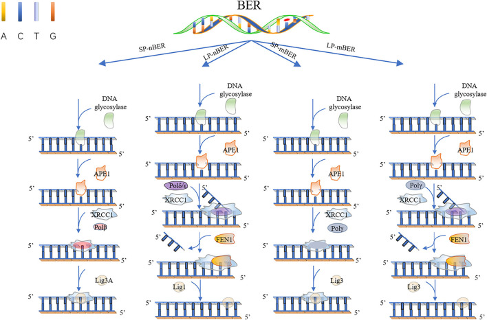
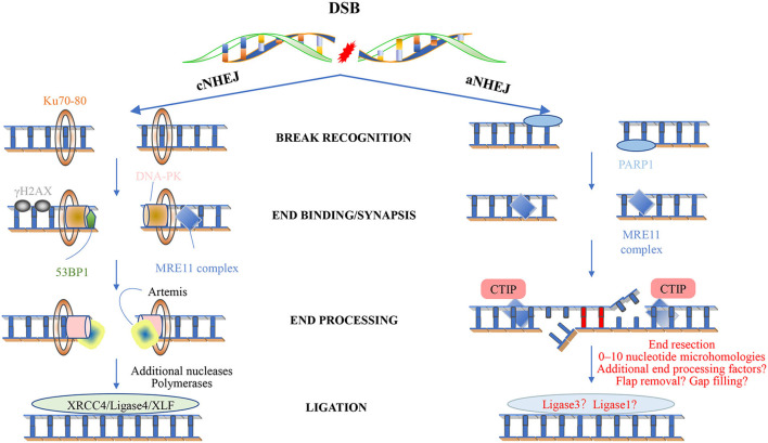
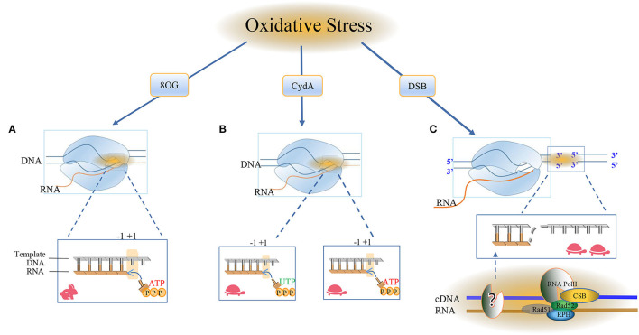
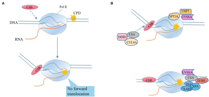
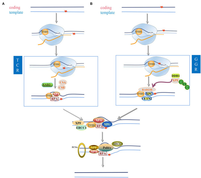
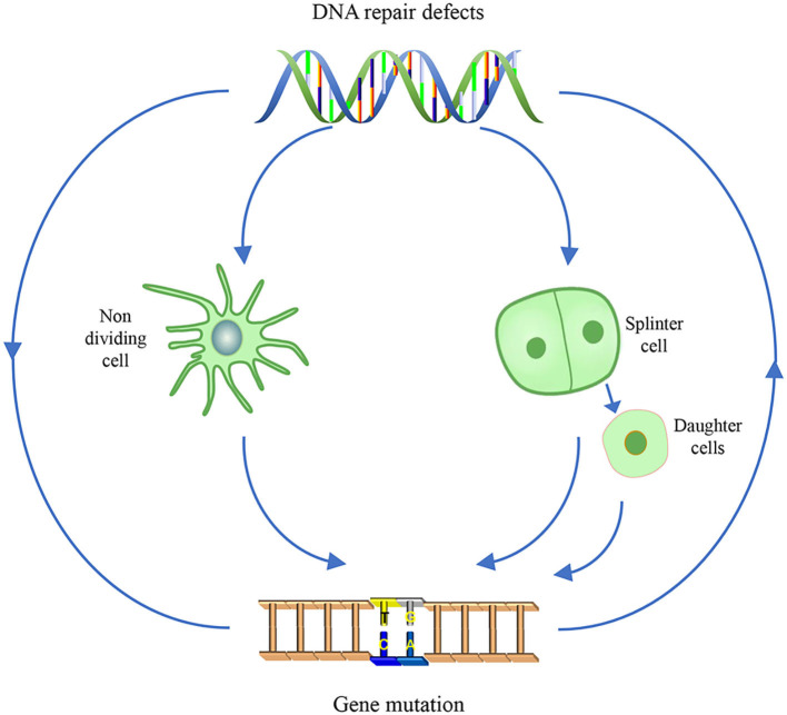
Most of the neurodegenerative diseases and aging are associated with reactive oxygen species (ROS) or other intracellular damaging agents that challenge the genome integrity of the neurons. As most of the mature neurons stay in G0/G1 phase, replication-uncoupled DNA repair pathways including BER, NER, SSBR, and NHEJ, are pivotal, efficient, and economic mechanisms to maintain genomic stability without reactivating cell cycle. In these progresses, polymerases are prominent, not only because they are responsible for both sensing and repairing damages, but also for their more diversified roles depending on the cell cycle phase and damage types. In this review, we summarized recent knowledge on the structural and biochemical properties of distinct polymerases, including DNA and RNA polymerases, which are known to be expressed and active in nervous system; the biological relevance of these polymerases and their interactors with neuronal degeneration would be most graphically illustrated by the neurological abnormalities observed in patients with hereditary diseases associated with defects in DNA repair; furthermore, the vicious cycle of the trinucleotide repeat (TNR) and impaired DNA repair pathway is also discussed. Unraveling the mechanisms and contextual basis of the role of the polymerases in DNA damage response and repair will promote our understanding about how long-lived postmitotic cells cope with DNA lesions, and why disrupted DNA repair contributes to disease origin, despite the diversity of mutations in genes. This knowledge may lead to new insight into the development of targeted intervention for neurodegenerative diseases.

Keywords: DNA polymerase; DNA repair pathway; RNA polymerase; neurodegenerative diseases; postmitotic cells.


Conflict of interest statement

The authors declare that the research was conducted in the absence of any commercial or financial relationships that could be construed as a potential conflict of interest.

Figures

Figure 1
Figure 1
Base excision repair. In short patch nuclear BER (SP-nBER), DNA glycosylase can cut the chemical bond between nucleotide bases and ribose to release a complete DNA phosphoribose chain. This process will form an purine or pyrimidine (AP) site. APE1 cleaves the 5'position of the phosphodiester chain at the AP site. In this way, a 3'-hydroxyl group and a 5'-basic deoxyribose phosphate group appear on the DNA strand. When 3'-hydroxyl and 5'-deoxyribose phosphate (dRP) are present, the SP-BER pathway continues, where DNA polymerase β (Polβ) removes 5'-dRP and inserts a new nucleotide to fill the gap, and then X-ray repairs the complex of cross-complementary protein 1 (XRCC1) and DNA ligase 3 (LIG3) to close the cut. In long patch nuclear BER (LP-nBER), the 5' end is not a Pol β substrate. In this pathway, 2–10 nucleotides at the 3'-end are replaced and removed from the DNA backbone, and the new nucleotide chain and petals shape endonuclease 1 (FEN1) complexes, and base complementary pairing is performed under the action of POL (δ or ε). The final ligation step is performed by LIG1. Mitochondrial BER (mtBER) includes short patch BER (SP-mBER) and long patch BER (LP-mBER). SP-mBER is initiated by a specific DNA glycosylase, which recognizes modified or inappropriate bases and cleaves N-glycosidic bonds to produce abasic site. The resulting AP site is processed by AP endonuclease, resulting in a strand break with a 3'-hydroxyl end and a 5'-(dRP) residue. Then, the mitochondrial DNA polymerase pol γ fills in the single-nucleotide gap for repair. In addition to polymerase activity, pol γ has 3'−5' exonuclease and 5'dRP lyase activities. Therefore, when mtBER is initiated by a monofunctional DNA glycosylase, the 5'-dRP part produced when the AP endonuclease cleaves the strand can be removed by the 5'-dRP lyase function of pol γ. Finally, the resulting nick is sealed by DNA ligase. mtBER can also be performed LP-mBER, which involves the incorporation of 2–12 nucleotides during the repair synthesis process. The LP-mBER treatment of DNA damage causes the DNA strands to be exposed as a part of a single-stranded overhang or flap structure. These flap structures are recognized and cleaved by flap endonuclease 1 (FEN-1), which is an essential enzyme for nuclear LP-mBER, and then ligated by DNA ligase.
Figure 2
Figure 2
Types of DNA damage and corresponding repair pathways. Endogenous and exogenous factors can induce different types of DNA damage. Here, we show their DDR mechanisms and cell proliferation status. The nerve cell model in the figure represents non-dividing cells, and the other model represents dividing cells. While LP-nBER, TLS and HRR exist in dividing cells, SP-nBER, LP-mBER, and SP-mBER exist in non-dividing cells. By contrast, MMR, A-NHEJ, and C-NHEJ exist not only in non-dividing cells but also in dividing cells. Here also shows DNA Pols related to different DNA damage repair pathways and diseases related to different types of damage. It is worth noting that Polγ only plays a role in mtBER. ALS, amyotrophic lateral sclerosis; FA, Fanconi syndrome; FAP, familial adenomatous polyposis; FTD, frontotemporal dementia; HD, Huntington's disease; HIM, hyper-IgM syndrome; HNPCC, hereditary non-polyposis colorectal cancer; PD, Parkinson's disease; SCA, spinocerebellar ataxia.
Figure 3
Figure 3
Non-homologous end joining. Classical NHEJ (cNHEJ) is triggered by the binding of a Ku heterodimer to the fragmented DNA end and provides a scaffold for the recruitment of other factors, including DNA-PKC, XRCC4 ligase IV-XLF, Artemis, and DNA polymerase. The Mre11 complex is loaded to the end of the DNA and can recruit ATM. The histone variant H2AX is phosphorylated to form γ-H2AX, which is located on both sides of the fracture. This in turn promotes the recruitment of other factors, leading to the assembly of large multi-protein complexes that may play a role in disrupting the signal, repairing and keeping DNA ends together, and minimizing the chance of abnormal rearrangements. cNHEJ requires additional enzymes to prepare the DNA-attached ends. One of the enzymes is Artemis, and once DNA-PK stimulates the endonuclease activity in Artemis, it will hit the development clamp. The last step of cNHEJ involves binding the DNA ends through the DNA ligase IV/XRCC4/XLF complex. In short, cNHEJ is a repair process in which the ends of DSBs are directly linked by DNA ligase, which does not rely on homologous DNA sequences. The Ku protein (Ku70/Ku80) complex recognizes and binds to the end of DSBs, and the Ku-DNA complex recruits DNA-dependent protein kinase catalytic subunits (DNA-PKcs) to activate its kinase activity, phosphorylate itself to initiate the NHEJ pathway, and attract the recombinase Artemis Join to process the DNA ends, and then summon the XRCC4-DNAligase4-XLF complex to promote the ligation of the DNA ends. Less is known about the mechanism of alternative NHEJ (aNHEJ). Although PARP1 can interact with free DNA ends, is associated with DNA damage induction, and can interact with ATM, the Mre11 complex seems to play an important role. However, the mechanism of its action is still unclear. Recent studies have shown that aNHEJ also occurs in cells that can activate other repair pathways. Future research will definitely learn more about this mechanism and its genome editing potential.
Figure 4
Figure 4
RNA Pol II bypasses the oxidative damage nucleotide addition cycle. The damage caused by oxidation here includes 8oxoG, CydA, and DSBs, and it is obvious that different cell types use different repair pathways at different rates. (A) Most of the oxidative modifications on bases can be bypassed by RNA Pol II. RNA Pol II bypasses the repair mechanism of oxidative damage and incorporates nucleotides into RNA. The RNA and Pol II form a ternary complex, in which the template DNA and the newly-born RNA form the core transcription bubble of Pol II. During the transcript extension process, RNA synthesis and the forward movement of Pol II will pass through the “Brown ratchet” coupling of translocation mechanisms. OG is one of the markers of oxidative stress. Pol II turns on the rapid transcription bypass, and DNA glycosylase triggers the release of specific DNA enzymes to identify and ablate damage and is used by BER in neuronal cells. Repair, the formation of ATP ready to pair with the base in the active site, the translocation mechanism creates a new RNA 3'end in the free +1 position in the active template, and RNA Pol II occupies the polymerase active site. (B) CydA is the damage in CydU that can cause Pol II transcription stagnation in human cells, opening a slow damage bypass, and polymerase is added to the UTP downstream of the CydA lesion and the DNA is pushed into the active position of RNA polymerase. Incorrectly adding AMP residue on the opposite side of the base of the downstream template, Pol II will translocate at the +1 position and slow down the subsequent elongation. (C) The surge of oxidative DNA damage puts too much pressure on the BER system and leads to DSB. Actively transcribed genes use transcription-coupled homologous recombination (TC-HR). RNA Pol II stagnates in the lesion, and the DNA-RNA hybrid structure recruits CSB. Then, the interaction between RNA Pol II and CSB initiates TC-HR and provides a scaffold for HR factors, such as Rad 52 and Rad 51C, which directly interact with CSB. There is a DNA polymerase upstream of the lesion site, which reverse-transcribes the template strand. The RNA polymerase II (Pol) in thermodynamics occupies the Pol active site. In the post-translocation state, elongation, nucleotide incorporation occurs through the “Bronen ratchet” site and reset Pol II to the pre-translocation state +1 (rabbit: fast, turtle: slow).
Figure 5
Figure 5
RNA polymerase II bypasses the transcriptional blocking damage cyclobutane pyrimidine dimer (CPD) triggers TC-NER repair. (A) CSB recognizes stagnant Pol II and binds to its upstream DNA, and uses its ATPase activity to push Pol II forward by translocating from 3'to 5'on the transcription template strand. CSB can move Pol II to natural pause sites and smaller lesions, but cannot push Pol II to larger TBLs, such as CPD (“stagnation”). The mechanism of detecting whether the captured Pol II can still translocate forward by pulling the DNA squeezed upstream can distinguish between the stagnant focus and the naturally suspended Pol II. (B) Transcription-coupled nucleotide excision repair model (TC-NER), from the arrest of RNA polymerase II (Pol II) to lesion excision and gap-filling DNA synthesis. After Pol II encounters TBL, its translocation activity induces the strong bending of upstream DNA and the tighter combination of CSB and Pol II, triggering transcription-coupled nucleotide excision repair (TC-NER). Chromatin remodeling agent stimulates the recruitment of CSB. CSA, DNA damage-binding protein 1 (DDB1) and cullin 4a (CUL4A)—RBX1 ubiquitin E3 ligase form the CRL4CSA complex, which is recruited to the lesion by CSB and recognizes damage signals after activation-NER starts. Ultraviolet-stimulated scaffold protein A (UVSSA) and ubiquitin C-terminal hydrolase 7 (USP7) are also recruited to the lesion, promoted by the chromatin remodeling subunit SPT16, and stably bind to CSA. CSB is ubiquitinated by CRL4CSA, but this is counteracted by USP7-mediated deubiquitylation to prevent CSB degradation. The transcription factor TFIIH is recruited through the interaction of its p62 subunit with UVSSA. TFIIH uses its 5'−3' XPD helicase to translocate forward on the DNA until it is blocked by the lesion, which may stimulate Pol II backtracking. XPA was confirmed to bind to TFIIH to recruit structure-specific endonucleases ERCC1-XPF and XPG. Cut the DNA 5' and 3'of the lesion, respectively, and release the 22–30 nucleotide long DNA oligomers containing the lesion. The resulting gap is filled by DNA synthesis, recruiting proliferating cell nuclear antigen (PCNA), replication factor C (RFC), and DNA polymerase δ, ε, and finally sealed by DNA ligase 1 or XRCC1-DNA ligase.
Figure 6
Figure 6
Nucleotide excision repair. In eukaryotic cells exposed to ultraviolet radiation, two different nucleotide excision repair (NER) modes are activated: (I) transcription-coupled nucleotide excision repair) (TC-NER) and (II) global genome Nucleotide excision repair (GG-NER), which participates in the recognition of twisted DNA and determines preferences related to space and time. NER helps eliminate spiral twisting damage, including cyclobutane-pyrimidine dimers (CPD), 6-4 photoproducts (6-4PPs), and other bulky adducts; therefore, it maintains the stability of the genome. The predictive influence of NER subpathway on the coding or template damaged chain of actively transcribed genes. (A,B) are located on the template strand (A) or coding strand (B) of the active gene to repair bulky lesions. If the lesion is located on the template strand (A), it is read by RNAPII during the transcription process, and the lesion will cause the RNAPII complex to stall. CSA and CSB proteins are sensors of stalled RNAPII and recruit the transcription complex TFIIH to the lesion. The helicase activity XPB and XPD of the TFIIH complex open the chromatin around the lesion. XPA and RPA stabilize the open structure of chromatin. Endonuclease, ERCC1-XPF in 5'and XPG in 3'cut the damaged chain. Then, the gap is filled by DNA repair polymerase and ligase. If the lesion is located on the coding strand (B) and therefore cannot be read by the RNAPII complex, the lesion will not interfere with the synthesis of the enzyme and the gene will be transcribed. The lesion can be identified by the XPC complex in the future. TFIIH opens a denatured bubble of about 30 nucleotides around the lesion. The complex of XPC, RAD23B, and CETN2 can directly bind to the opposite DNA strand, where the spirally twisted lesion accumulates XPC is recruited to these damaged sites only after the UV-DDB (ultraviolet-damaged DNA-binding protein) complex binds. XPC complex is a disease-binding protein in global genome repair (GG-NER). After the DNA helix is partially opened, RPA (replication protein A) is added to the complex, which then helps in damage verification. XPA is best combined with single-stranded DNA (double-stranded DNA) structure, while RPA can only be observed in the ssDNA (single-stranded DNA region. This is the second NER subpathway. After the lesion recognition step, GG-NER and TC-NER are the same in the air bubbles before the incision, XPA has been shown to be located on the 5'side of the lesion. XPF-ERCC1 catalyzes the 5'incision, and XPG is responsible for the 3'incision around the lesion. ERCC1 is polyubiquitinated at the K33 site, which can be It is removed by USP45 (ubiquitin-specific peptidase 45). In TC-NER, when RNA polymerase II stays on the lesion during the transcription extension process, the lesion is recognized. RNA polymerase II (RNAPII) is due to active genes. The damage in the transcription chain (TS) stalls and attracts NER enzymes, RNAPII (RNA polymerase II), and CSB (Cockayne syndrome B protein) to further repair the protein. It can be deubiquitinated by USP7 to keep recruited in the lesion. Proliferating cell nuclear antigen (PCNA) is loaded to the 5'end of the DNA. PCNA interacts with XPA and XPF to stimulate their activity. The DNA region containing 22–30 nucleotides is excised from the complex DNA with TFIIH and then slowly released from TFIIH, bound by RPA or degraded by nuclease. During the nicking step, XPG is simultaneously ubiquitinated by CRL4Cdt2 and then degraded in the 26S proteasome. DNA synthesis is catalyzed by DNA polymerase δ/ε. Precise coordination of ubiquitin-mediated RNAPII removal after transcriptional blockade.
Figure 7
Figure 7
The vicious cycle of gene mutation and impaired DNA repair pathway in TNR diseases. For non-dividing cells, gene mutation disrupting the DNA repair pathway is displayed as cell senescence and apoptosis, that is the outcome of the TNR diseases. For dividing cells, the aberrant DNA repair pathway in turn exacerbates the TNR expansion. In somatic cells, the gene mutation will be passed on and accumulate as the cells proliferate; In germ cells, the aggravated gene mutation is inherited by the offspring, which will exhibit more severe TNA disease symptom in neurons system and aggravated mutagenicity in somatic cells.

Similar articles

Cited by

References

    1. Abbotts R., Wilson D. M. (2017). Coordination of DNA single strand break repair. Free Radic. Bio Med. 107, 228–244. 10.1016/j.freeradbiomed.2016.11.039 - DOI - PMC - PubMed
    1. Abramzon Y. A., Fratta P., Traynor B. J., Chia R. (2020). The overlapping genetics of amyotrophic lateral sclerosis and frontotemporal dementia. Front. Neurosci. 14, 42–51. 10.3389/fnins.2020.00042 - DOI - PMC - PubMed
    1. Ahel I., Rass U., El-Khamisy S. F., Katyal S., Clements P. M., McKinnon P. J. (2006). The neurodegenerative disease protein aprataxin resolves abortive DNA ligation intermediates. Nature 443, 713–716. 10.1038/nature05164 - DOI - PubMed
    1. Akbari M., Peña-Diaz J., Andersen S., Liabakk N. B., Otterlei M., Krokan H. E. (2009). Extracts of proliferating and non-proliferating human cells display different base excision pathways and repair fidelity. DNA Repair 8, 834–843. 10.1016/j.dnarep.2009.04.002 - DOI - PubMed
    1. Almaguer-Mederos L. E., Jorge-Sainz Y., Almaguer-Gotay D., Aguilera-Rodríguez R., Rodríguez-Labrada R., Velázquez-Pérez L., et al. . (2020). One-carbon metabolism factor MTHFR variant is associated with saccade latency in Spinocerebellar Ataxia type 2. J. Neurol. Sci. 409, 116586. 10.1016/j.jns.2019.116586 - DOI - PubMed