Review
. 2022 Jul 1:20:574-597.
doi: 10.1016/j.bioactmat.2022.06.011. eCollection 2023 Feb.

Integrated gradient tissue-engineered osteochondral scaffolds: Challenges, current efforts and future perspectives

Affiliations
Review

Integrated gradient tissue-engineered osteochondral scaffolds: Challenges, current efforts and future perspectives

Xiaolian Niu et al. Bioact Mater. .

Abstract

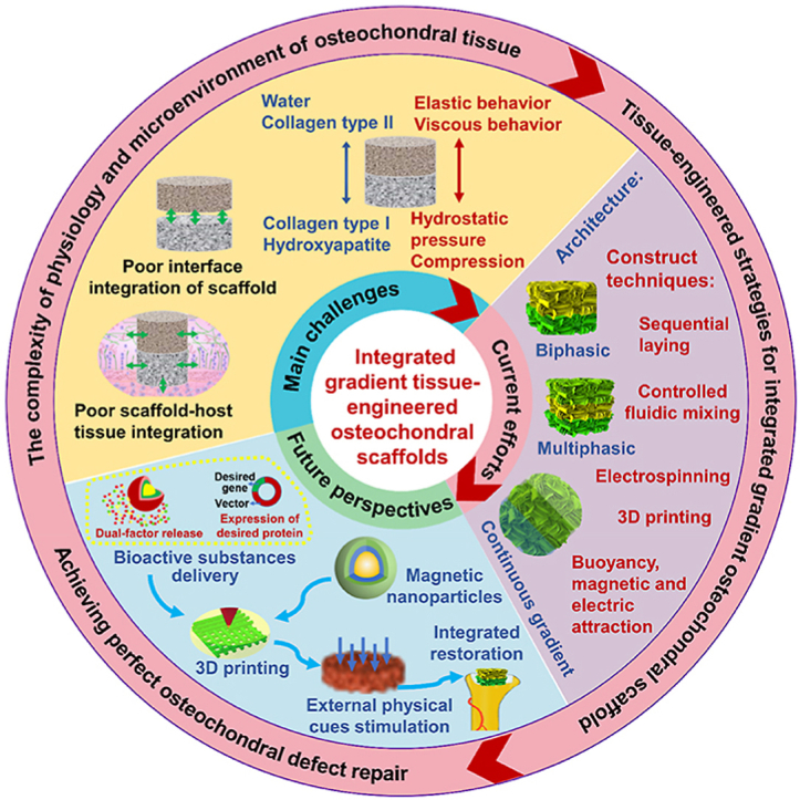
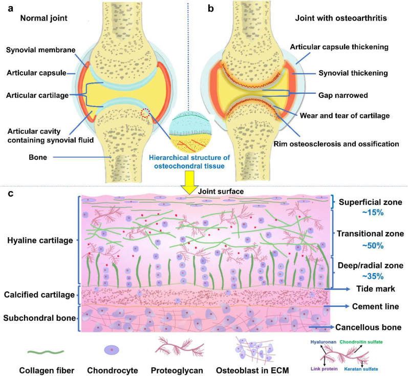
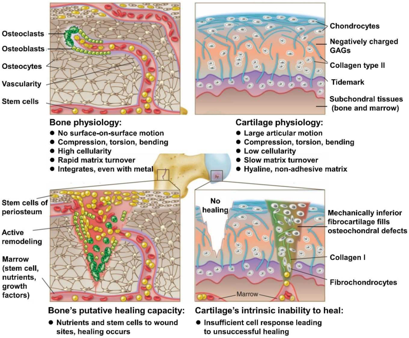
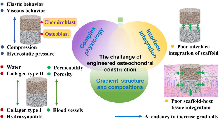
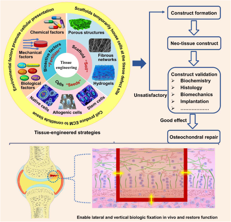
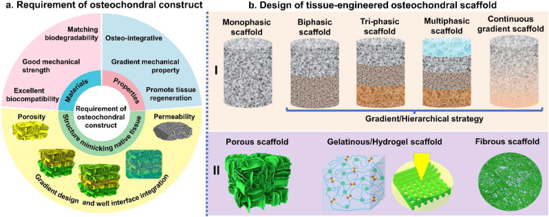
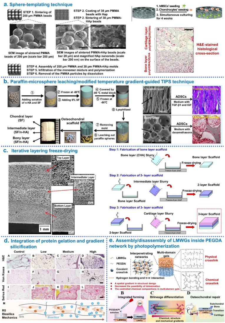
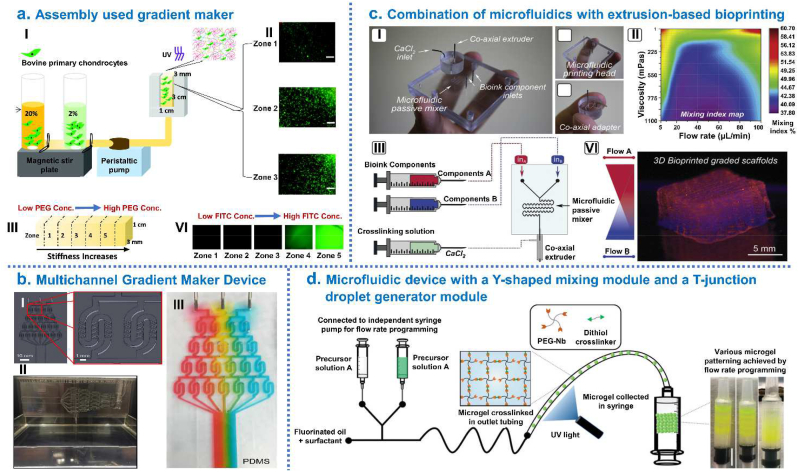
The osteochondral defect repair has been most extensively studied due to the rising demand for new therapies to diseases such as osteoarthritis. Tissue engineering has been proposed as a promising strategy to meet the demand of simultaneous regeneration of both cartilage and subchondral bone by constructing integrated gradient tissue-engineered osteochondral scaffold (IGTEOS). This review brought forward the main challenges of establishing a satisfactory IGTEOS from the perspectives of the complexity of physiology and microenvironment of osteochondral tissue, and the limitations of obtaining the desired and required scaffold. Then, we comprehensively discussed and summarized the current tissue-engineered efforts to resolve the above challenges, including architecture strategies, fabrication techniques and in vitro/in vivo evaluation methods of the IGTEOS. Especially, we highlighted the advantages and limitations of various fabrication techniques of IGTEOS, and common cases of IGTEOS application. Finally, based on the above challenges and current research progress, we analyzed in details the future perspectives of tissue-engineered osteochondral construct, so as to achieve the perfect reconstruction of the cartilaginous and osseous layers of osteochondral tissue simultaneously. This comprehensive and instructive review could provide deep insights into our current understanding of IGTEOS.

Keywords: Evaluation; Fabrication techniques; Integrated gradient tissue-engineered osteochondral scaffold (IGTEOS); Osteochondral tissue engineering; Tissue-engineered strategies.


Conflict of interest statement

There are no conflicts of interest to declare.

Figures

Image 1
Graphical abstract
Fig. 1
Fig. 1
The schematic diagram of (a) normal joint, (b) diseased joint, and (c) osteochondral unit including cartilage, calcified cartilage and subchondral bone.
Fig. 2
Fig. 2
The schematic diagram illustrated the difference in the physiologic environment and healing capacities of cartilage and bone tissue. Reproduced with permission [5]: copyright 2012, AAAS.
Fig. 3
Fig. 3
Challenges of making an integrated gradients tissue-engineered osteochondral construct for clinical use, including complex physiology, interface integration, and gradient structure and composition.
Fig. 4
Fig. 4
The building process of a tissue-engineered osteochondral construct: tissue-engineered osteochondral strategies usually resort to the combination of innovative biomaterials, cells and signal molecule, aiming to recapitulate the biological, physical and functional features of the native osteochondral unit; after repeated evaluation and validation, such biomimicking constructs could then be implanted into a damaged osteochondral region, where they will assist tissue repair, promote regenerative responses and facilitate the functional recovery of the joint.
Fig. 5
Fig. 5
Tissue-engineered strategies of osteochondral scaffold. (a) Requirement of integrated gradient tissue-engineered osteochondral construct: the materials of osteochondral scaffold should have matching biodegradability, good mechanical strength and excellent biocompatibility; the structure of osteochondral scaffold should mimic native tissue, including suitable pore sizes and porosity, gradient design and well interface integration; some properties of osteochondral scaffold are essential, such as good osseointegration, gradient mechanical property and improved tissue regeneration. (b) Schematic diagram of design of tissue-engineered osteochondral scaffold in vitro: I) Scaffold strategies could be classified according to the number of layers and gradient properties of the designs; II) Micromorphology of osteochondral scaffold.
Fig. 6
Fig. 6
Integrated hierarchical osteochondral scaffold was designed by sequential layering techniques. (a) Steps of the sphere-templating technique to fabricate an integrated bi-layered scaffold and in vitro cell study. (b) Schematic diagrams of the process for preparing integrated osteochondral scaffolds by combining paraffin-sphere leaching with a modified temperature gradient-guided thermal-induced phase separation (TIPS) technique. (c) The “iterative layering freeze-drying” fabrication process diagram to fabricate collagen-based scaffold with a seamlessly integrated layer structure for osteochondral defect repair. (d) The process of generate seamlessly integrated bilayer hydrogel for osteochondral defect repair by simultaneously polymerizing two layers using a one-pot method. (e) The mechanism of formation of the multi-domain gel and its great potential for osteochondral regeneration through controlling chemical, structural, and mechanical properties of each gel domain. Reproduced with permission: (a) [29], copyright 2013, Wiley; (b) [68], copyright 2014, ACS; (c) [54], copyright 2014, Elsevier; (d) [81], copyright 2019, Wiley; (e) [85], copyright 2021, Elsevier.
Fig. 7
Fig. 7
Integrated hierarchical osteochondral scaffold was designed by 3D printing techniques. (a) Preparation of biphasic scaffold by 3D stereolithography printer: GelMA-PEGDA as primary ink, TGF-β1/PLGA NPs loaded into the top layer and nHA loaded into the bottom layer of osteochondral scaffold. (b) Fabrication of a bio-inspired multilayer osteochondral scaffold that consisted of the PCL and HA/PCL microspheres via selective laser sintering layer-by-layer process. (c) Fabrication of biohybrid gradient PNT scaffolds by thermal-assisted extrusion 3D printing for repair of osteochondral defect. (d) 3D printing gradient PACG-GelMA hydrogel scaffolds assisted with a low-temperature receiver: the bioactive Mn2+ are loaded into the top cartilage layer while the BG is incorporated into the bottom subchondral bone layer. (e) Fabrication process of tissue-engineered osteochondral scaffolds through integrate fused deposition modeling 3D printing with a casting technique. (f) Fabrication of biphasic HA/PCL scaffolds by multi-nozzle 3D printer. Reproduced with permission: (a) [43], copyright 2019, Elsevier; (b) [7], copyright 2017, Elsevier; (c) [42], copyright 2018, Wiley; (d) [86], copyright 2019, Wiley; (e) [72], copyright 2019, Elsevier; (f) [71], copyright 2021, Springer.
Fig. 8
Fig. 8
Integrated hierarchical osteochondral scaffold was designed by controlled fluidic mixing techniques. (a) Gradient hydrogel fabrication and characterization: (I) Schematic representation of gradient maker assembly used to make gradient hydrogel which is bulk polymerized after the prepolymer solution is mixed with bovine primary chondrocytes; (II) Cell viability within selected zones of the gradient hydrogel on day; (III) Compressive modulus from zone 1 to zone 5 in gradient hydrogel; (VI) Dual-gradient hydrogel with biochemical model protein (FITC tagged Bovine Serum Albumin-BSA) encapsulation could also be achieved. (b) Development of Multichannel Gradient Maker Device (MGMD): (I) Solidworks 3D computer-aided design software used to design the MGMD to facilitate chaotic mixing in channels; (II) PDMS MGMDs were generated using 3D printed molds and a syringe pump was used to flow solutions through MGMD channels; (III) Colored dyes were mixed with 70% glycerol and pumped through the MGMD to visually display gradient generation. (c) Combination of microfluidics with extrusion-based bioprinting and instructive bioinks to produce graded scaffolds: (I) Microfluidic extrusion system composed of the microfluidic printing head and the co-axial adapter; (II) Mixing index heatmap; (III) schematically shown how to 3D bioprint graded scaffolds. (d) Microgel production procedure using a microfluidic device with a Y-shaped mixing module and a T-junction droplet generator module. Right side photograph showing examples of microgel patterning. Reproduced with permission: (a) [164], copyright 2018, Mary Ann Liebert; (b) [176], copyright 2019, Elsevier; (c) [162], copyright 2019, IOP; (d) [165], copyright 2020, Wiley.
Fig. 9
Fig. 9
Integrated hierarchical osteochondral scaffold was designed by buoyancy, magnetic attraction and electric attraction techniques. (a) Growth factor gradients for osteochondral tissue engineering: I) Osteochondral tissue, engineered using of hMSC-laden GelMA hydrogels, with buoyancy used to form a morphogen gradient of BMP-2 complexed with heparin methacrylate (HepMA); II) Alizarin Red S staining revealed localized mineral deposition at one end of the tissue; III) Alcian Blue staining revealed tissue-wide staining for glycosaminoglycans, a component of both cartilage and bone [116]. (b) Engineering osteochondral tissue using magnetically-aligned glycosylated SPIONs: (I) SPIONs were conjugated with heparin to produce a glycosylated corona that could efficiently sequester and release growth factors; (II) An external magnetic field was used to field-align glycosylated SPIONs in a hMSC-laden agarose hydrogel, which was thermally gelled and cultured for 28 days to generate robust osteochondral constructs comprising both bone and cartilage tissue; (III) Finite element modeling of the magnetic field strength and distribution; (VI) The key mineralization protein osteopontin (red), which were present specifically at the bone end of the tissue [117]. (c) Using electric field migration to fabricate silk nanofiber hydrogels with gradients and the control of cell differentiation [118]. Reproduced with permission: (a) [116], copyright 2019, Wiley; (b) [117], copyright 2018, Elsevier; (c) [118], copyright 2020, Springer.

References

    1. Mahmoudian A., Lohmander L.S., Mobasheri A., Englund M., Luyten F.P. Early-stage symptomatic osteoarthritis of the knee - time for action. Nat. Rev. Rheumatol. 2021;17:621–632. - PubMed
    1. Wang X., Lin J., Li Z., Ma Y., Zhang X., He Q., Wu Q., Yan Y., Wei W., Yao X., Li C., Li W., Xie S., Hu Y., Zhang S., Hong Y., Li X., Chen W., Duan W., Ouyang H. Identification of an ultrathin osteochondral interface tissue with specific nanostructure at the human knee joint. Nano Lett. 2022;22:2309–2319. - PubMed
    1. Khanarian N.T., Haney N.M., Burga R.A., Lu H.H. A functional agarose-hydroxyapatite scaffold for osteochondral interface regeneration. Biomaterials. 2012;33:5247–5258. - PMC - PubMed
    1. Heinegård D., Saxne T. The role of the cartilage matrix in osteoarthritis. Nat. Rev. Rheumatol. 2011;7:50–56. - PubMed
    1. Huey D.J., Hu J.C., Athanasiou K.A. Unlike bone, cartilage regeneration remains elusive. Science. 2012;338:917–921. - PMC - PubMed

LinkOut - more resources