. 2022 Apr 2:17:526-541.
doi: 10.1016/j.bioactmat.2022.03.015. eCollection 2022 Nov.

Engineering microparticles based on solidified stem cell secretome with an augmented pro-angiogenic factor portfolio for therapeutic angiogenesis

Affiliations

Engineering microparticles based on solidified stem cell secretome with an augmented pro-angiogenic factor portfolio for therapeutic angiogenesis

Thomas Später et al. Bioact Mater. .

Abstract

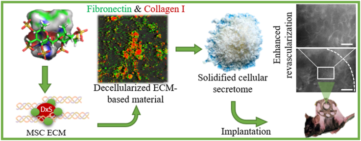
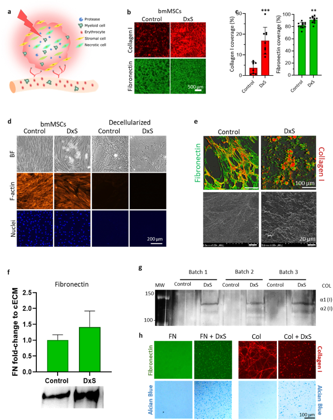
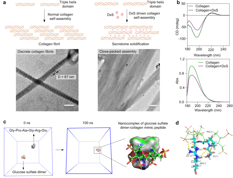
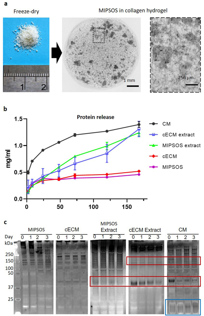
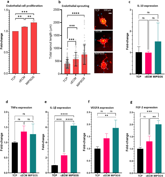
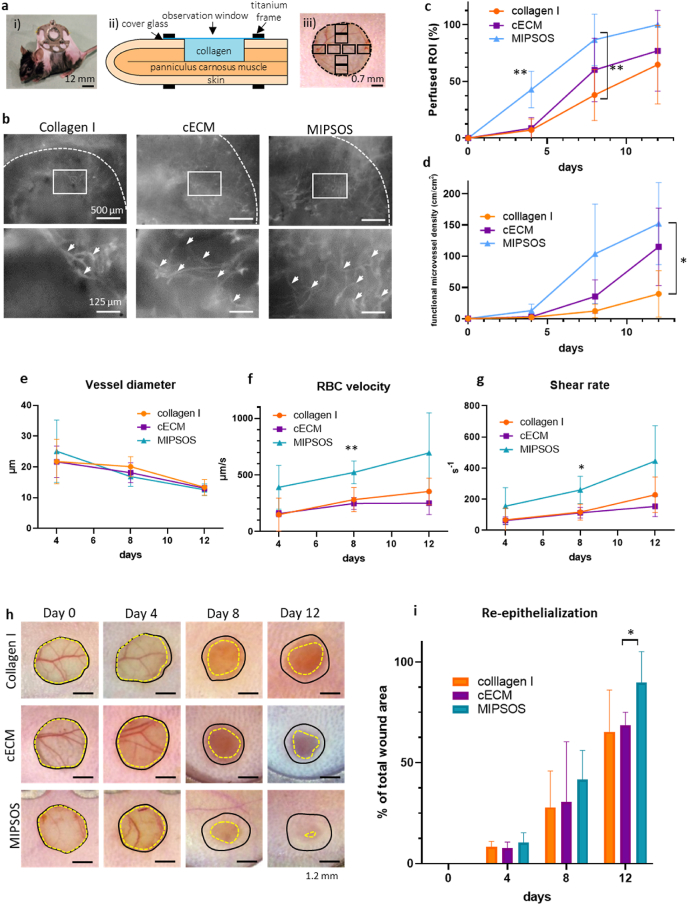
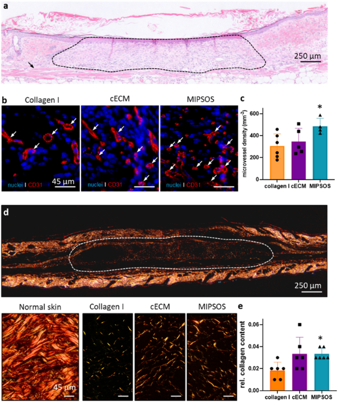
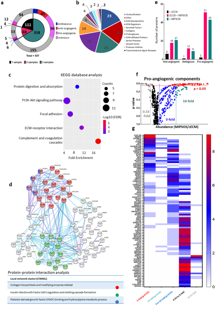
Tissue (re)vascularization strategies face various challenges, as therapeutic cells do not survive long enough in situ, while the administration of pro-angiogenic factors is hampered by fast clearance and insufficient ability to emulate complex spatiotemporal signaling. Here, we propose to address these limitations by engineering a functional biomaterial capable of capturing and concentrating the pro-angiogenic activities of mesenchymal stem cells (MSCs). In particular, dextran sulfate, a high molecular weight sulfated glucose polymer, supplemented to MSC cultures, interacts with MSC-derived extracellular matrix (ECM) components and facilitates their co-assembly and accumulation in the pericellular space. Upon decellularization, the resulting dextran sulfate-ECM hybrid material can be processed into MIcroparticles of SOlidified Secretome (MIPSOS). The insoluble format of MIPSOS protects protein components from degradation, while facilitating their sustained release. Proteomic analysis demonstrates that MIPSOS are highly enriched in pro-angiogenic factors, resulting in an enhanced pro-angiogenic bioactivity when compared to naïve MSC-derived ECM (cECM). Consequently, intravital microscopy of full-thickness skin wounds treated with MIPSOS demonstrates accelerated revascularization and healing, far superior to the therapeutic potential of cECM. Hence, the microparticle-based solidified stem cell secretome provides a promising platform to address major limitations of current therapeutic angiogenesis approaches.

Keywords: Dextran sulfate; Extracellular matrix; Mesenchymal stem cells; Poly-electrolyte-driven co-assembly; Therapeutic angiogenesis; Wound healing.


Conflict of interest statement

AB and MA would like to declare the filing of a PCT patent application (WO 2020/228733 A1).

Figures

Image 1
Graphical abstract
Fig. 1
Fig. 1
Dextran sulfate-mediated deposition and assembly of MSC-derived ECM. (a) Schematic of an ischemic injury. Impaired vascularization of tissue leads to necrotic cells, which expand from the center into the periphery. This promotes an inflammatory response characterized by myeloid cell infiltration and enhanced proteolytic activity. (b) Immunofluorescence staining of collagen type I (red) and fibronectin (green) deposited by cultured bone marrow-derived mesenchymal stem cells (bmMSC) on day 6 in the absence (control) and presence of dextran sulfate (DxS). Bar = 500 μm (c) Corresponding quantification of stained surface area per field of view. (d) Decellularized ECM. Complete decellularization of assembled ECM is evidenced by the absence of F-actin and nuclear (DAPI) staining. BF, bright-field microscopy. Bar = 200 μm. (e) Collagen type I and fibronectin immunostaining and topography of decellularized ECM. Confocal microscopy (top) and scanning electron microscopy (bottom) of decellularized ECM of control and dextran sulfate-treated bmMSC cultures (DxS). Bar = 100 μm (top) or 20 μm (bottom). (f) Western blot analysis of fibronectin (FN) in decellularized control and dextran sulfate-supplemented cultures (DxS) and densitometric analysis of immune-reactive bands (n = 3). (g) Silver stained SDS-PAGE of pepsin-digested samples of control ECM and ECM deposited in the presence of dextran sulfate (DxS) showing collagen type I (COL) α1 and α2 bands. (h) Purified fibronectin (FN) or collagen type I (Col) were incubated with dextran sulfate (100 μg/ml) and deposited structures were visualized by immunocytochemistry. Deposited dextran sulfate was visualized by staining with Alcian Blue at a pH 2.5 before immunostaining. *, p < 0.05; **, p < 0.01; ***, p < 0.001.
Fig. 2
Fig. 2
Co-assembly of collagen and dextran sulfate and molecular motifs. (a) Transmission electron microscopy imaging of the effect of dextran sulfate (DxS, 10 μg/ml) on collagen type I fibrillogenesis, visualized by negative staining with phosphotungstic acid (Top row: graphic depiction of potential co-assembly process). Scale bar = 250 nm. (b) Analysis of the effect of dextran sulfate on the molecular assembly of collagen type I (0.2 mg/ml) based on far-UV and circular dichroism (CD). (c and d) Simulation of the molecular dynamics of dextran sulfate collagen type I interaction using a glucose sulfate dimer and a typical collagen type I hexapeptide Gly-Pro-Ala-Gly-Arg-Glu, respectively.
Fig. 3
Fig. 3
Stability of ECM-based microparticles. (a) Hydrogel construct with MIPSOS. Lyophilized cECM and MIPSOS resembled insoluble microparticles and could be encapsulated in a collagen type I scaffold. (b) Protein release profiles. Protein content released from cECM and MIPSOS entrapped in collagen hydrogels into the supernatant was estimated using BCA assay. (c) Protein degradation profiles. The microparticle formulation of MIPSOS and cECM protected protein contents from degradation, as compared to corresponding urea-solubilized extracts and MSC-conditioned medium (CM), when incubated at 37 °C for up to 3 days in PBS. Red and blue squares: Bands showing a marked decrease and increase, respectively, in density over time.
Fig. 4
Fig. 4
Bioactivity of ECM-based microparticles. (a) Endothelial cell proliferation on various substrates. HUVECs were seeded on TCP (control), cECM or MIPSOS and cultured for 3 days. Relative cell numbers were determined by CCK8 assay. (b) Endothelial sprouting assay on various substrates. The assay and quantification of cumulative sprout length/spheroid was performed using endothelial spheroids in a collagen hydrogel construct cast on tissue culture polystyrene (TCP), cECM or MIPSOS. Asterisks indicate tips of the formed sprouts. Scale bar = 100 μm. PCR for (c) IL-10, (d) TNFα, (e) IL-1β, (f) VEGFA and (g) FGF-2 expressed by THP-1 derived macrophages seeded on various substrates for 144 h.**, p < 0.01, ***, p < 0.001; ****, p < 0.0001.
Fig. 5
Fig. 5
MIPSOS promoted revascularization in vivo. (a) Representative illustration of the experimental in vivo model, showing (i) a C57BL/6 mouse with dorsal skinfold chamber, (ii) cross-sectional schematic of skinfold chamber set-up and (iii) a full-thickness skin defect with indicated regions of interest (ROIs, black squares), which were analyzed by means of intravital fluorescence microscopy. (b) Intravital fluorescence microscopy of ingrowing perfused micro-vessels after intravenous injection of 5% FITC-labeled dextran 150,000 in full-thickness skin defects (Ø 3 mm) on day 8 after implantation. Dashed lines indicate the initial defect margins. White arrow heads point to vessel ramifications. (c) Percentage of perfused ROIs and (d) functional microvessel density (in cm/cm2) in wounds. (e) Diameter (μm) and (f) centerline red blood cell (RBC) velocity (μm/s) of 40 randomly selected microvessels. (g) Wall shear rate (y, in s−1) calculated from values obtained in (e) and (f) by means of the Newtonian definition y = 8 × v/d. (h) Wound re-epithelialization of full-thickness skin defects over a period of 12 days. Black lines indicate initial defect margins. Yellow dashed lines indicate progression of re-epithelialization. (i) Quantification of wound area over time * p < 0.05; **p < 0.01.
Fig. 6
Fig. 6
MIPSOS promoted wound healing. (a) Representative HE-stained histological sections of full-thickness mouse skin wounds treated with MIPSOS particle-laden collagen gel on day 12. The area of hydrogel implant is indicated by dashed line. Bar = 250 μm. (b) Representative images of CD31-stained microvessels in skin defects on day 12; nuclei were counterstained with DAPI. Bar = 45 μm. (c) Quantified microvessel density in mm−2. (d) Polarized light microscopy of Sirius red-stained sections of normal skin as well as healed skin defects on day 12. Bar = 250 μm (top) or 45 μm (bottom). (e) Corresponding quantification of relative collagen signal (implant/skin) on day 12 after implantation. *, p < 0.05, compared to collagen I.
Fig. 7
Fig. 7
Pro-angiogenic components are enriched in MIPSOS. (a) Pie diagram visualizing distribution of identified secretome proteins from three biological replicates according to their reported role in angiogenesis. (b) A total of 133 proteins with MIPSOS/cECM abundance ratio >3 folds were further classified based on Matrisome Project and Panther classification system. (c) Using KEGG database in DAVID bioinformatics, 5 pathways (FDR <0.05) were identified and organized by false discovery rate FDR value as well as protein counts. (d) Protein-protein interaction (PPI) based on STRING Network analysis (Confidence score threshold at 0.7 (high)) highlights significant protein interaction networks. Proteins are represented as nodes of different colors. (e) Number of proteins identified in all three biological replicates (from a) categorized based on their enrichment (3-fold difference) in cECM (

Similar articles

Cited by

References

    1. Manuel G.E., Johnson T., Liu D. Therapeutic angiogenesis of exosomes for ischemic stroke. Int. J. Physiol. Pathophysiol. Pharmacol. 2017;9:188–191. - PMC - PubMed
    1. Eming S.A., Martin P., Tomic-Canic M. Wound repair and regeneration: mechanisms, signaling, and translation. Sci. Transl. Med. 2014;6:265sr6. doi: 10.1126/SCITRANSLMED.3009337. - DOI - PMC - PubMed
    1. Zhao L., Johnson T., Liu D. Therapeutic angiogenesis of adipose-derived stem cells for ischemic diseases. Stem Cell Res. Ther. 2017;8 doi: 10.1186/s13287-017-0578-2. - DOI - PMC - PubMed
    1. Wei Z., Volkova E., Blatchley M.R., Gerecht S. Hydrogel vehicles for sequential delivery of protein drugs to promote vascular regeneration. Adv. Drug Deliv. Rev. 2019;149–150:95–106. doi: 10.1016/j.addr.2019.08.005. - DOI - PMC - PubMed
    1. Johnson T., Zhao L., Manuel G., Taylor H., Liu D. Approaches to therapeutic angiogenesis for ischemic heart disease. J. Mol. Med. 2019;97:141–151. doi: 10.1007/s00109-018-1729-3. - DOI - PMC - PubMed

LinkOut - more resources