. 2022 Jul 1:9:885241.
doi: 10.3389/fsurg.2022.885241. eCollection 2022.

Vancomycin Containing PDLLA and PLGA/β-TCP Inhibit Biofilm Formation but Do Not Stimulate Osteogenic Transformation of Human Mesenchymal Stem Cells

Affiliations

Vancomycin Containing PDLLA and PLGA/β-TCP Inhibit Biofilm Formation but Do Not Stimulate Osteogenic Transformation of Human Mesenchymal Stem Cells

Berna Kankilic et al. Front Surg. .

Abstract

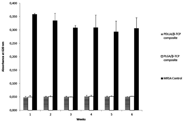
Aims: Chronic osteomyelitis, including implant-related prosthetic joint infection, is extremely difficult to cure. We develop vancomycin containing release systems from poly(d,l-lactide) (PDLLA) and poly(d,l-lactide-co-glycolide) (PLGA) composites with beta-tricalcium phosphate (β-TCP) to treat methicillin-resistant Staphylococcus aureus osteomyelitis. We ask whether vancomycin containing PDLLA/β-TCP and PLGA/β-TCP composites will prevent early biofilm formation, allow cell proliferation and osteogenic differentiation, and stimulate osteogenic signaling molecules in the absence of an osteogenic medium.

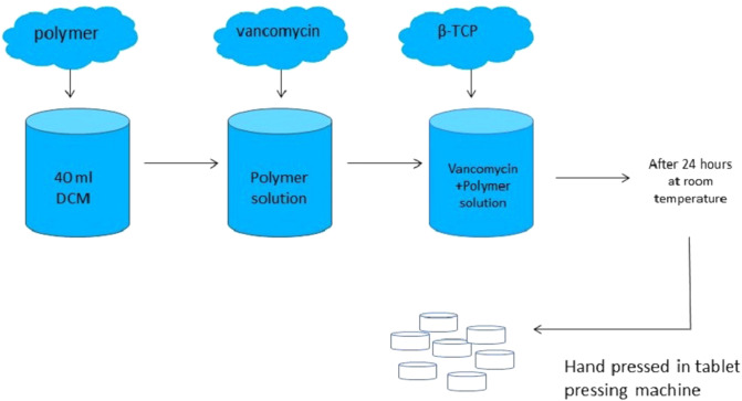
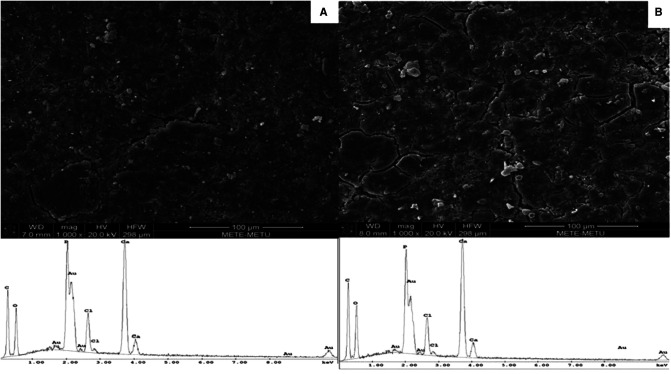
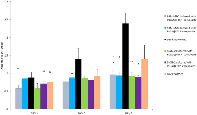
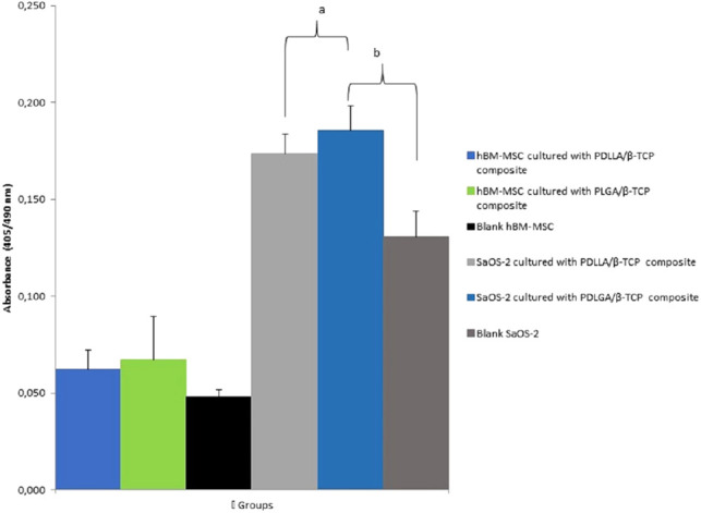
Methods: Composites were produced and characterized with scanning electron microscopy. In vitro vancomycin release was assessed for 6 weeks. Biofilm prevention was calculated by crystal violet staining. Human bone marrow-derived mesenchymal stem cells (hBM-MSCs) and osteosarcoma cell (SaOS-2) proliferation and differentiation were assessed with water soluble tetrazolium salt and alkaline phosphatase (ALP) staining. Real-time quantitative polymerase chain reaction defined osteogenic signaling molecules for hBM-MSCs.

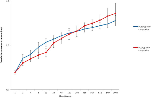
Results: Totally, 3.1 ± 0.2 mg and 3.4 ± 0.4 mg vancomycin released from PDLLA/β-TCP and the PLGA/β-TCP composites, respectively, and inhibited early biofilm formation. hBM-MSCs and SaOS-2 cells proliferated on the composites and stimulated ALP activity of cells. Runt-related transcription factor 2 (RUNX2) and SRY-Box transcription Factor 9 (SOX9) expressions were, however, lower with composites when compared with control.

Conclusion: Vancomycin containing PDLLA/β-TCP and PLGA/β-TCP composites inhibited early biofilm formation and proliferated and differentiated hBM-MSCs and SaOS-2 cells, but osteogenesis-related RUNX2 and SOX9 transcription factors were not strongly expressed in the absence of an osteogenic medium for 14 days.

Keywords: PDLLA; PLGA; biofilm; bone signaling molecules; vancomycin; β-TCP.


Conflict of interest statement

One or more of the authors (BK, EB, PK, and FK) has received funding from the Republic of Turkey Ministry of Science, Industry and Technology SANTEZ Programme Project No: 00817.STZ.2011-1.

Figures

Figure 1
Figure 1
The design of the composites.
Figure 2
Figure 2
SEM micrographs (1,000× magnification) and EDS spectra of vancomycin containing (A) PDLLA/β-TCP and (B) PLGA/β-TCP composites. PDLLA, poly(d,l-lactide); PLGA, poly(d,l-lactide-co-glycolide); β-TCP, beta-tricalcium phosphate; EDS, energy-dispersive spectrometry; SEM, scanning electron microscope.
Figure 3
Figure 3
FTIR spectra of (A) vancomycin, (B) β-TCP, (C) PDLLA, (D) vancomycin containing PDLLA/β-TCP composite, (E) PLGA, and (F) vancomycin containing PLGA/β-TCP composite. PDLLA, poly(d,l-lactide); PLGA, poly(d,l-lactide-co-glycolide); β-TCP, beta-tricalcium phosphate.
Figure 4
Figure 4
Cumulative released vancomycin amount from the composites in 6 weeks. PDLLA, poly(d,l-lactide); PLGA, poly(d,l-lactide-co-glycolide); β-TCP, beta-tricalcium phosphate.
Figure 5
Figure 5
Absorbance results of bacterial control, PDLLA/β-TCP and PLGA/β-TCP composites at 620 nm (weeks are defining the time point of drug release media collected from release studies). PDLLA, poly(d,l-lactide); PLGA, poly(d,l-lactide-co-glycolide); β-TCP, beta-tricalcium phosphate; MRSA, methicillin-resistant Staphylococcus aureus.
Figure 6
Figure 6
Proliferation of the MSC and SaOS-2 cells in days 1, 3, and 7 according to the absorbance of WST-1 at 450 nm. PDLLA, poly(d,l-lactide); PLGA, poly(d,l-lactide-co-glycolide); β-TCP, beta-tricalcium phosphate; SaOS-2, osteosarcoma cell; hBM-MSC, human bone marrow-derived mesenchymal stem cell; WST, water soluble tetrazolium salt.
Figure 7
Figure 7
Alkaline phosphatase (ALP) activity of the groups at day 21 according to their absorbances at 405 nm. PDLLA, poly(d,l-lactide); PLGA, poly(d,l-lactide-co-glycolide); β-TCP, beta-tricalcium phosphate; SaOS-2, osteosarcoma cell; hBM-MSC, human bone marrow-derived mesenchymal stem cell.

Similar articles

Cited by

References

    1. Birt MC, Anderson DW, Toby EB, Wang J. Osteomyelitis: recent advances in pathophysiology and therapeutic strategies. J Orthop. (2017) 14(1):45–52. 10.1016/jor.2016.10.004 - DOI - PMC - PubMed
    1. Inzana JA, Schwarz EM, Kates SL, Awad HA. Biomaterials approaches to treating implant-associated osteomyelitis. Biomaterials. (2016) 81:58–71. 10.1016/j.biomaterials.2015.12.012 - DOI - PMC - PubMed
    1. Choi HR, Bedair H. Mortality following revision total knee arthroplasty: a matched cohort study of septic versus aseptic revisions. J Arthroplasty. (2014) 29:1216–8. 10.1016/j.arth.2013.11.026 - DOI - PubMed
    1. Kamath AF, Ong KL, Lau E, Chan V, Vail TP, Rubash HE, et al. Quantifying the burden of revision total joint arthroplasty for periprosthetic infection. J Arthroplasty. (2015) 30(9):1492–7. 10.1016/j.arth.2015.03.035 - DOI - PubMed
    1. Taha M, Abdelbary H, Ross FP, Carli AV. New innovations in the treatment of PJI and biofilms—clinical and preclinical topics. Curr Rev Musculoskelet Med. (2018) 11(3):380–8. 10.1007/s12178-018-9500-5 - DOI - PMC - PubMed