. 2022 Jul 1:13:911250.
doi: 10.3389/fphar.2022.911250. eCollection 2022.

Hepatoprotective Efficacy and Interventional Mechanism of Qijia Rougan Decoction in Liver Fibrosis

Affiliations

Hepatoprotective Efficacy and Interventional Mechanism of Qijia Rougan Decoction in Liver Fibrosis

Xiao-Feng Chen et al. Front Pharmacol. .

Abstract

Liver fibrosis is a leading contributor to chronic liver diseases such as cirrhosis and liver cancer, which pose a serious health threat worldwide, and there are no effective drugs to treat it. Qijia Rougan decoction was modified from Sanjiasan, a traditional Chinese medicine (TCM) described in the "Wenyilun" manuscript. Qijia Rougan decoction possesses hepatoprotective and antifibrotic effects for clinical applications. However, its underlying mechanisms remain largely unknown. In this study, fibrotic rats induced by carbon tetrachloride (CCl4) were treated with two doses of Qijia Rougan decoction. Histopathological and serum biochemical analyses were carried out to assess liver structure and function, respectively. High-performance liquid chromatography (HPLC) coupled with mass spectrometry (MS) was performed to identify bioactive compositions in Qijia Rougan decoction. Transcriptome analysis using mRNA-sequencing (mRNA-Seq) was used to explore the underlying mechanisms and verified by quantitative real-time polymerase chain reaction (qRT-PCR) and Western blotting. Qijia Rougan decoction significantly attenuated CCl4-induced hepatic fibrotic injury, supported by promoted liver function and improved liver fibrosis. Eight main representative components originating from raw materials in the Qijia Rougan decoction were found to possess an antifibrotic role. Mechanistically, Qijia Rougan decoction regulated biological processes such as oxidation-reduction, fatty acid metabolism, cell adhesion, and transforming growth factor beta (TGFβ) signaling. We determined that Qijia Rougan decoction reversed the expression of inflammatory cytokines and inhibited the activation of fibrosis-related TGFβ signaling. It also reversed the deterioration of liver structure and function in rats induced by CCl4. Overall, Qijia Rougan decoction significantly mediated metabolism-associated processes, inhibited inflammatory reactions, and repressed fibrosis-related TGFβ signaling, which prevented liver fibrosis deterioration. Our study deepens our understanding of TCM in the diagnosis and treatment of liver fibrosis.

Keywords: inflammation; liver fibrosis; qijia rougan decoction; transcriptomics; transforming growth factor beta.


Conflict of interest statement

The authors declare that the research was conducted in the absence of any commercial or financial relationships that could be construed as a potential conflict of interest.

Figures

FIGURE 1
FIGURE 1
Total ion current chromatograms (TICCs) of the Qijia Rougan decoction (QJ). (A) TICCs of the negative (ESI−) ionization modes of the QJ; (B) TICCs of the positive (ESI+) ionization modes of the QJ.
FIGURE 2
FIGURE 2
Detection of liver index and serum biochemical indicators of rats in four groups. (A) Body weight (n = 8–16); (B) Liver weight (n = 8–16); (C) Liver index = Liver weight/Body weight X 100% (n = 8–16); (D) ALT: alanine aminotransferase (n = 8–16); (E) AST: aspartate aminotransferase (n = 8–16); (F) TBIL: total bilirubin (n = 8–16); (G) HA: hyaluronic acid (n = 8–10); (H) PC III: type III procollagen (n = 8–10); (I) LN: laminin (n = 8–10). *p < 0.05 and ***p < 0.001. ns, not significant.
FIGURE 3
FIGURE 3
Qijia Rougan decoction protects the rat liver from CCl4-induced injury. (A) Hematoxylin and eosin (H&E, scale bar = 100 µm) staining was performed to detect liver structure in rats; (B) Masson’s staining (scale bar = 100 µm) was performed to detect liver fibrosis in rats; (C) Quantification of liver fibrosis in four groups (n = 8–10); (D,E) qRT-PCR was used to analyze Col1a1 and Timp1 mRNA levels in rats (n = 6–12); (F,G) Immunohistochemical (IHC) staining was performed to detect the level of α-SMA and collagen I in the rat liver. *p < 0.05 and ***p < 0.001.
FIGURE 4
FIGURE 4
DEG analysis of the mRNA-Seq. (A–D) Volcano plot of differentially expressed genes in Qijia Rougan decoction–treated rats with fibrosis. (A) Model vs. Control; (B) QJ-L vs. Model; (C) QJ-H vs. Model; (D) QJ-H vs. QJ-L; (E) Numbers of the overlapping genes in different comparisons (Venn); (F) Expression heatmap in the four experimental groups; (G) Corrplot correlation heat map analyses between the four groups.
FIGURE 5
FIGURE 5
DEG Target analysis of QJ-H. (A) Overlapping DEGs between Model vs. Control-down and QJ-H vs. Model-up (Venn); (B) Overlapping DEGs between Model vs. Control-up and QJ-H vs. Model-down (Venn); (C) Heatmap for hierarchical cluster analysis of DEGs between Control, Model, and QJ-H.
FIGURE 6
FIGURE 6
GO and KEGG enrichment analysis of the DEG target of QJ-H. (A) GO analysis histogram of the 1590 overlapping upregulated DEGs between Model vs. Control-down and QJ-H vs. Model-up; (B) GO analysis histogram of the 1444 overlapping downregulated DEGs between Model vs. Control-up and QJ-H vs. Model-down; (C) KEGG enrichment analysis of the 1590 overlapping upregulated DEGs between Model vs. Control-down and QJ-H vs. Model-up; (D) KEGG enrichment analysis of the 1444 overlapping downregulated DEGs between Model vs. Control-up and QJ-H vs. Model-down.
FIGURE 7
FIGURE 7
Expression level and Gene Set Enrichment Analysis (GSEA) analysis of TGF-beta signaling. (A–C) Bulk RNA-seq analysis of the expression level of Tgfb1, Tgfb2, and Tgfb3 (n = 3); (D–F) Gene Set Enrichment Analysis (GSEA) analysis of TGF-beta signaling. NES, normalized enrichment score; positive and negative NESs indicate higher and lower expression, respectively. *p < 0.05, **p < 0.01, and ***p < 0.001. ns, not significant.
FIGURE 8
FIGURE 8
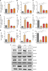
Validation of the expression of mRNA and proteins. (A–F) qRT-PCR validations of related genes (n = 4); (A) Tgfb1: transforming growth factor beta 1; (B) Tgfb2: transforming growth factor, beta 2; (C) Tgfb3: transforming growth factor beta 3; (D) Tgfb1i1: transforming growth factor beta 1–induced transcript 1; (E) Cxcl1: C-X-C motif chemokine ligand 1; (F) Cxcl12: C-X-C motif chemokine ligand 12; (G–I) Bulk RNA-seq analysis of the expression level of Tgfb1i1, Cxcl1, and Cxcl12 (n = 3); (J) Representative Western blotting was performed to detect the levels of p-Smad3, Smad3, p-Smad2, Smad2, TGF-βR1, TGF-βR2, and TGF-β.

Similar articles

Cited by

References

    1. Ali M., Khan T., Fatima K., Ali Q. U. A., Ovais M., Khalil A. T., et al. (2018). Selected Hepatoprotective Herbal Medicines: Evidence from Ethnomedicinal Applications, Animal Models, and Possible Mechanism of Actions. Phytother. Res. 32 (2), 199–215. 10.1002/ptr.5957 - DOI - PMC - PubMed
    1. Chen S., Zou L., Li L., Wu T. (2013). The Protective Effect of Glycyrrhetinic Acid on Carbon Tetrachloride-Induced Chronic Liver Fibrosis in Mice via Upregulation of Nrf2. PLoS One 8 (1), e53662. 10.1371/journal.pone.0053662 - DOI - PMC - PubMed
    1. Chen H., Cai J., Wang J., Qiu Y., Jiang C., Wang Y., et al. (2021). Targeting Nestin+ Hepatic Stellate Cells Ameliorates Liver Fibrosis by Facilitating TβRI Degradation. J. Hepatol. 74 (5), 1176–1187. 10.1016/j.jhep.2020.11.016 - DOI - PubMed
    1. Dewidar B., Meyer C., Dooley S., Meindl-Beinker A. N. (2019). TGF-β in Hepatic Stellate Cell Activation and Liver Fibrogenesis-Updated 2019. Cells 8 (11), 1419. 10.3390/cells8111419 - DOI - PMC - PubMed
    1. Fabregat I., Moreno-Càceres J., Sánchez A., Dooley S., Dewidar B., Giannelli G., et al. (2016). TGF-β Signalling and Liver Disease. FEBS J. 283 (12), 2219–2232. 10.1111/febs.13665 - DOI - PubMed

LinkOut - more resources