. 2022 Jun 30:13:880258.
doi: 10.3389/fmicb.2022.880258. eCollection 2022.

In vitro Anti-Hantavirus Activity of Protein Kinase Inhibitor 8G1 Targeting AKT/mTOR/eIF4E Signaling Pathway

Affiliations

In vitro Anti-Hantavirus Activity of Protein Kinase Inhibitor 8G1 Targeting AKT/mTOR/eIF4E Signaling Pathway

Zhoupeng Li et al. Front Microbiol. .

Abstract

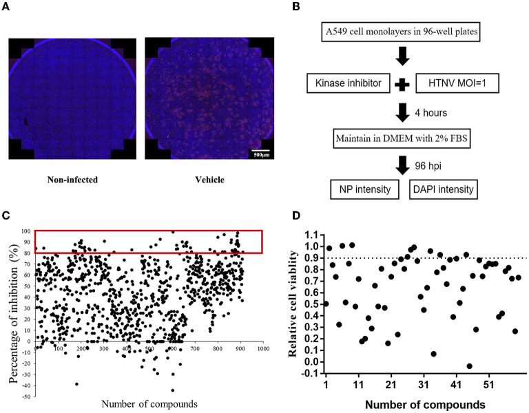
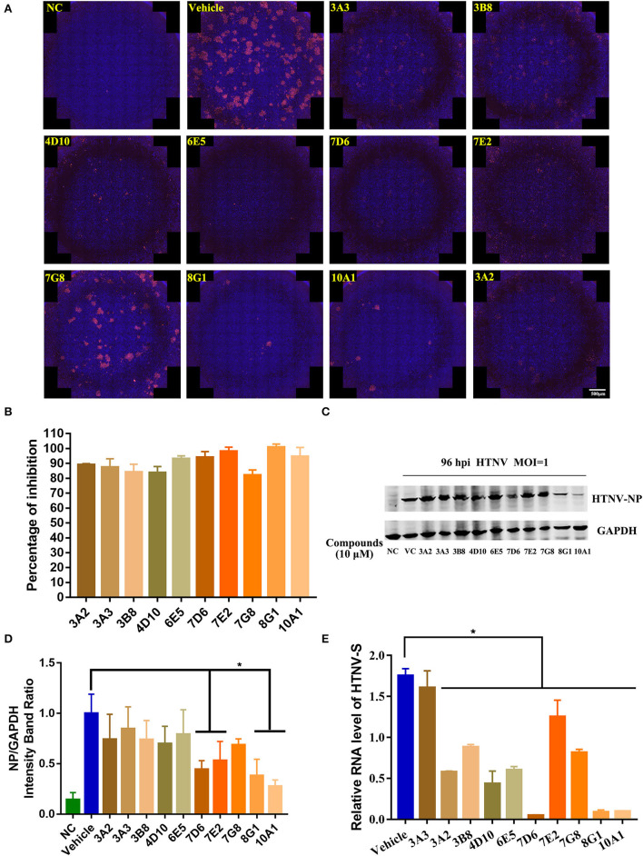
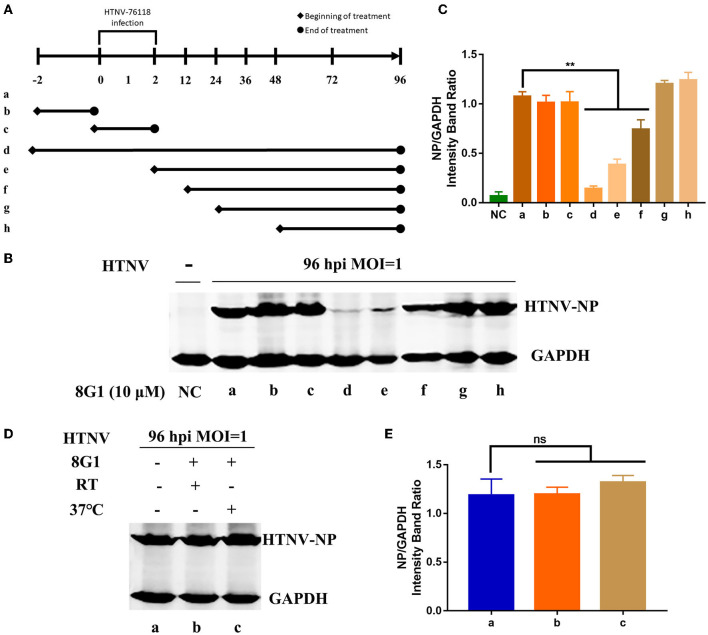
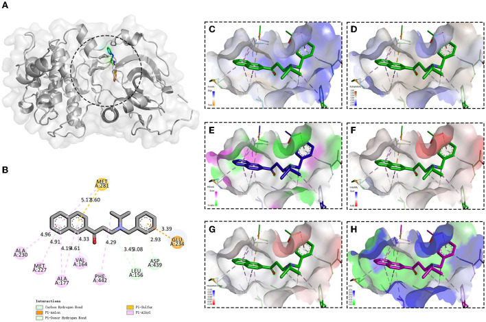
Hantaan virus (HTNV) is the main cause of hemorrhagic fever with renal syndrome (HFRS) around the world, which results in profound morbidity and mortality. However, there are currently no FDA-approved therapeutics or vaccines against HFRS. To find new anti-HTNV drugs, the inhibitory activity of 901 small molecule kinase inhibitors against HTNV is analyzed. Among these compounds, compound 8G1 inhibits HTNV with a relatively high inhibition rate and lower toxicity. The viral titer and nucleocapsid protein of HTNV are reduced after compound 8G1 treatment in a dose-dependent manner at concentrations ranging from 1 to 20 μM. In addition, the administration of compound 8G1 at the early stage of HTNV infection can inhibit the replication of HTNV. The molecular docking result reveals that compound 8G1 forms interactions with the key amino acid residues of serine/threonine-protein kinase B (Akt), which is responsible for the observed affinity. Then, the mammalian target of rapamycin (mTOR) and eukaryotic translation initiation factor 4E (eIF4E) signaling pathways are inhibited. Our results may help to design novel targets for therapeutic intervention against HTNV infection and to understand the anti-HTNV mechanism of protein kinase inhibitors.

Keywords: Hantaan virus; inhibitor; nucleocapsid protein; protein kinase; the mammalian target of rapamycin.


Conflict of interest statement

The authors declare that the research was conducted in the absence of any commercial or financial relationships that could be construed as a potential conflict of interest.

Figures

Figure 1
Figure 1
The screening of protein kinase inhibitors with anti-Hantaan virus (anti-HTNV) activity. (A) Observation of the anti-HTNV activity of kinase inhibitors by immunofluorescence assay (IFA), red staining for the expression of nucleocapsid protein (NP), and blue staining for the cell nuclei, scale bar = 500 μm. (B) Preliminary high-throughput screening of protein kinase inhibitors with anti-HTNV activity. (C) Among 901 protein kinase inhibitors, 60 could inhibit the HTNV more than 80%. (D) Among the above 60 protein kinase inhibitors, 10 compounds had no significant effect on cell viability.
Figure 2
Figure 2
Identification of potential kinase inhibitor candidates with in vitro anti-HTNV activity. (A) Immunofluorescence staining of HTNV nucleocapsid protein after the 10 protein kinase inhibitors treatment, NC, negative control, Vehicle: Vehicle-treated control, scale bar = 500 μm. (B) Inhibitory percentage of protein kinase inhibitors to virus infection based on the immunofluorescence staining. (C) The representative Western blot staining for HTNV NP protein in the negative control (NC) group, Vehicle-treated control (VC) group, and 10 μM 10 protein kinase inhibitors treatment groups. (D) The analyzed result of expression of HTNV nucleocapsid protein in the NC, VC, and 10 protein kinase inhibitors treatment groups in A549 cells post HTNV infection, *p < 0.05 vs. Vehicle, n = 3. (E) Inhibitory activity of protein kinase inhibitors to the expression of HTNV-S gene extracted from intracellular RNA, the mRNA expression level of HTNV-S gene was normalized to the respective β-actin and analyzed, *p < 0.05 vs. Vehicle, n = 3.
Figure 3
Figure 3
Evaluation of the selectivity index of three kinase inhibitor candidates. The chemical structure and dose-dependent inhibitory activity of (A) compound 7D6, (B) compound 8G1, and (C) compound 10A1 against HTNV at concentrations ranging from 1 to 20 μM. The half cytotoxicity concentration of (D) compound 7D6, (E) compound 8G1, and (F) compound 10A1 was measured at a concentration ranging from 1 to 100 μM, n = 3.
Figure 4
Figure 4
Compound 8G1 inhibited HTNV replication in A549 cell post-infection. (A) The representative Western blot staining for HTNV NP protein in the negative control (NC) group, Vehicle-treated control group, and compound 8G1 (1, 2.5, 5, 10, and 20 μM) treatment groups. (B) The analyzed result of expression of HTNV nucleocapsid protein in the NC, Vehicle, and compound 8G1 treatment groups in A549 cells post-HTNV infection, p < 0.05 vs. Vehicle, n = 3. (C) The representative imaging for the focus-forming unit of HTNV in the Vehicle group and compound 8G1 (1, 2.5, 5, 10, and 20 μM) treatment groups. (D) The viral titer of HTNV was analyzed in the Vehicle group and compound 8G1 treatment groups. **p < 0.01 vs. Vehicle, n = 3. (E) The expression of HTNV-S gene in the Vehicle group and compound 8G1 (1, 2.5, 5, 10, and 20 μM) groups, *p < 0.05 vs. Vehicle, n = 3.
Figure 5
Figure 5
Compound 8G1 reduced the expression of HTNV-NP proteins at the early stage of HTNV infection. (A) Different administration regimens of compound 8G1 treated the HTNV-infected A549 cells at an MOI of 1.0. Lane a: without compound 8G1 treatment; lane b: compound 8G1 treatment for 2 h pre-infection; lane c: compound 8G1 treatment for 2 h post-infection (hpi); lane d–h: treatment continues from 2 h pre-infection, 2, 12, 24, and 48 hpi to the 96 hpi time point. (B) The representative Western blot staining for HTNV NP protein in the groups with different administration regimens of compound 8G1. (C) The analyzed result of the expression of HTNV-NP protein in groups with different administration regimens of compound 8G1. **p < 0.01 vs. a, n = 3. (D) The representative Western blot staining for HTNV NP protein in the A549 cells without or in the presence of compound 8G1 at different temperatures. (E) The analyzed result of the expression of HTNV NP protein in control or compound 8G1 treatment groups at room temperature (RT) or 37°C.
Figure 6
Figure 6
Binding site and model of compound 8G1 with AKT. (A) Overview of the docked pose between the compound 8G1 and binding pocket to the active site of Akt. (B) The close view of the interaction between compound 8G1 and the active site of Akt. (C) The analyzed result of the aromatic ring edges or faces. (D) The analyzed result of the hydrophobicity. (E) The analyzed result of the hydrogen bond. (F) The analyzed result of the ionizability. (G) The analyzed result of the atomic charge, values less than −0.1 are mapped in red, and values larger than +0.1 are mapped in blue. (H) The analyzed result of the solvent accessibility surface, small values (green) correspond to buried residues, whereas large values (blue) correspond to exposed residues.
Figure 7
Figure 7
Effect of compound 8G1 to the protein kinase B (Akt)/mammalian target of rapamycin (mTOR) pathway. (A) The representative Western blot staining for phosphorylation of Akt and mTOR proteins in A549 cells. (B) The analyzed result of the expression of p(Ser473) Akt, *p < 0.05, #p < 0.05 vs. DMSO treated group, **p < 0.01 vs. HTNV group, n = 3. (C) The analyzed result of the expression of p-mTOR, **p < 0.01 vs. DMSO treated group, ***p < 0.005 vs. HTNV group, n = 3. (D) The analyzed result of the expression of HTNV-NP, **p < 0.001 vs. HTNV group, n = 3. (E) A549 cells were with treated 10 μM 8G1 or DMSO infected in the presence of inhibitors for 96 h before cells were harvested. The phosphorylation status of 4E-BP1 and elF4E was analyzed using antibodies against total 4E-BP1, p-4E-BP1 (Thr70), p-4E-BP1 (Ser65), p-4E-BP1 (Thr37/46), non-p-4E-BP1 (Thr46), p-eIF4E, and eIF4E. Total protein was isolated and analyzed by the Western blot using antibodies as shown. aA549 cells treated with solvent DMSO in the absence of HTNV infection; bA549 cells treated with compound 8G1 in the absence of HTNV infection; cA549 cells treated with the solvent DMSO in the context of HTNV infection; dA549 cells treated with compound 8G1 in the context of HTNV infection.

Similar articles

Cited by

References

    1. Avsic-Zupanc T., Saksida A., Korva M. (2019). Hantavirus infections. Clin. Microbiol. Infect. 21S, e6–e16. 10.1111/1469-0691.12291 - DOI - PubMed
    1. Engdahl T. B., Kuzmina N. A., Ronk A. J., Mire C. E., Hyde M. A., Kose N. (2021). Broad and potently neutralizing monoclonal antibodies isolated from human survivors of new world hantavirus infection. Cell Rep. 35:109086. 10.1016/j.celrep.2021.109086 - DOI - PMC - PubMed
    1. Fedeli C., Moreno H., Kunz S. (2020). The role of receptor tyrosine kinases in lassa virus cell entry. Viruses 12:80857. 10.3390/v12080857 - DOI - PMC - PubMed
    1. Hayashi Y., Watanabe H., Okudaira N., Otomo E., Endo S. (1981). The effects of butoctamide hydrogen succinate (bahs) on the sleep of the aged people (author's transl). No To Shinkei 33, 1243–1250. - PubMed
    1. Indrayanto G., Putra G. S., Suhud F. (2021). Validation of in-vitro bioassay methods: application in herbal drug research. Profiles Drug Subst. Excip. Relat. Methodol. 46, 273–307. 10.1016/bs.podrm.2020.07.005 - DOI - PubMed