. 2022 Sep 7;5(9):2960-2974.
doi: 10.1016/j.matt.2022.06.012. Epub 2022 Jul 11.

Inhalable dry powder mRNA vaccines based on extracellular vesicles

Affiliations

Inhalable dry powder mRNA vaccines based on extracellular vesicles

Kristen D Popowski et al. Matter. .

Abstract

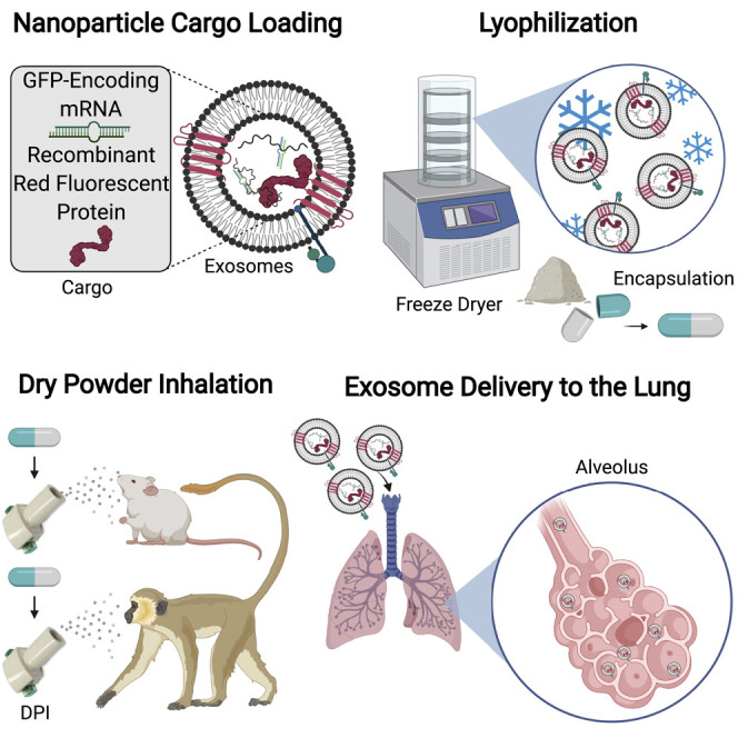
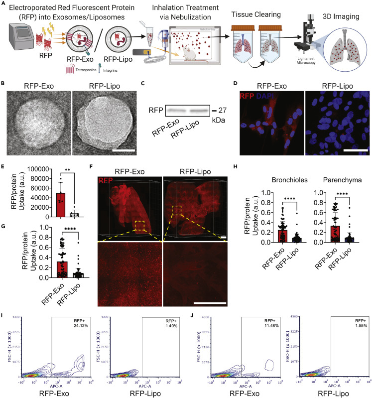
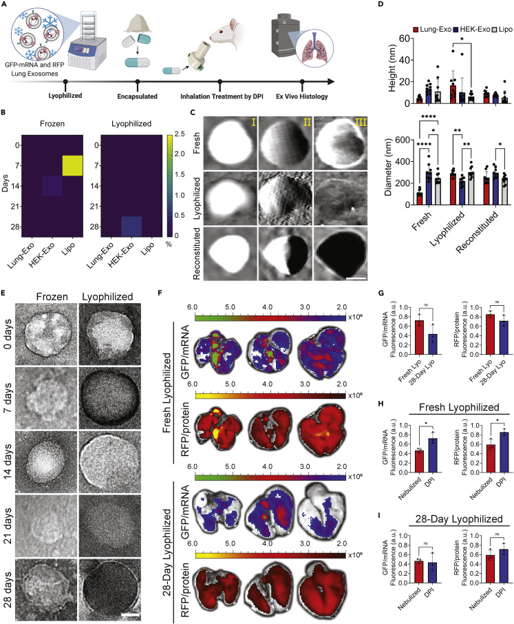
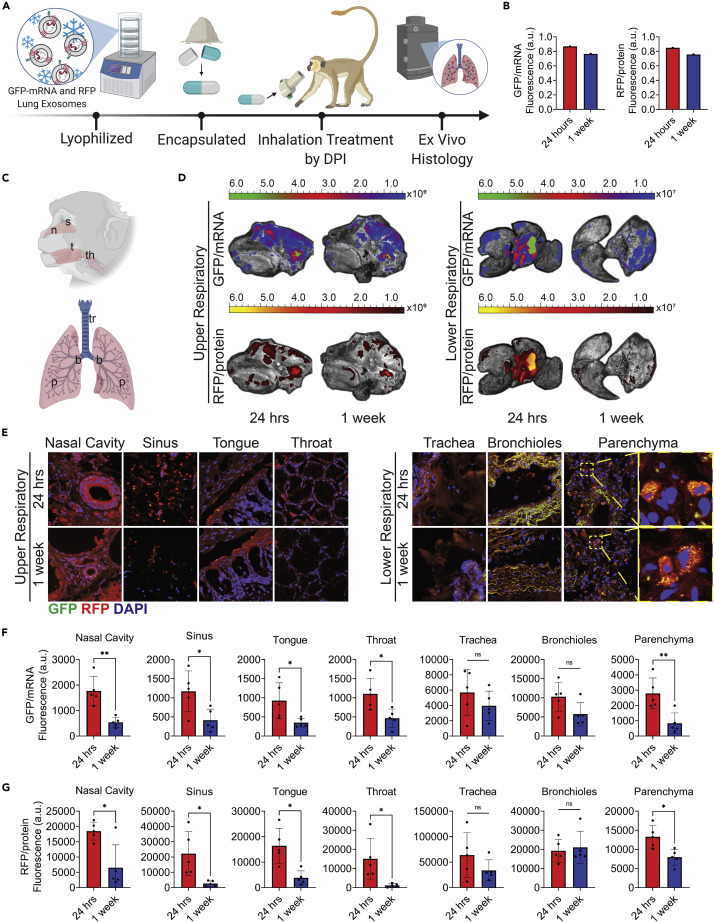
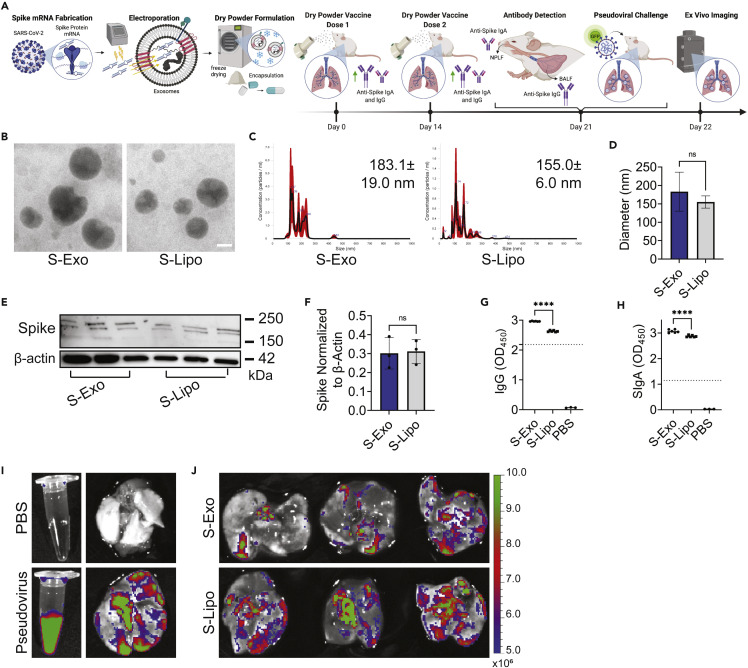
Respiratory diseases are a global burden, with millions of deaths attributed to pulmonary illnesses and dysfunctions. Therapeutics have been developed, but they present major limitations regarding pulmonary bioavailability and product stability. To circumvent such limitations, we developed room-temperature-stable inhalable lung-derived extracellular vesicles or exosomes (Lung-Exos) as mRNA and protein drug carriers. Compared with standard synthetic nanoparticle liposomes (Lipos), Lung-Exos exhibited superior distribution to the bronchioles and parenchyma and are deliverable to the lungs of rodents and nonhuman primates (NHPs) by dry powder inhalation. In a vaccine application, severe acute respiratory coronavirus 2 (SARS-CoV-2) spike (S) protein encoding mRNA-loaded Lung-Exos (S-Exos) elicited greater immunoglobulin G (IgG) and secretory IgA (SIgA) responses than its loaded liposome (S-Lipo) counterpart. Importantly, S-Exos remained functional at room-temperature storage for one month. Our results suggest that extracellular vesicles can serve as an inhaled mRNA drug-delivery system that is superior to synthetic liposomes.

Keywords: COVID-19; dry powder inhalation; exosomes; extracellular vesicles; lung; mRNA vaccine; nonhuman primate; spike protein.


Conflict of interest statement

North Carolina State University has filed a patent on the technologies related to this study. K.C. is an equity holder and consultant of Xsome Biotech, Inc. Xsome has entered an exclusive license agreement with North Carolina State University.

Figures

None
Graphical abstract
Figure 1
Figure 1
Fabrication and distribution of exosomes and liposomes (A) Schematic showing protein loading into lung-derived exosomes (RFP-Exos) and liposomes (RFP-Lipos), nebulization administration, ex vivo lung-tissue clearing, and 3D imaging by LSFM. Created with BioRender.com. (B) TEM images of RFP-Exos and RFP-Lipos; scale bar: 50 nm. (C) Immunoblot of RFP in exosome and liposome lysate. (D) Representative immunostaining images of lung parenchymal cells for RFP (red) and DAPI (blue); scale bar: 50 μm. (E) Quantification of RFP-Exo and RFP-Lipo pixel intensity normalized to nuclei in lung parenchymal cell images; n = 6 per group; data are represented as mean ± standard deviation. (F) LSFM images of cleared mouse lungs after RFP-Exo and RFP-Lipo nebulization; scale bar: 1,000 μm. (G) Quantification of the integrated density of RFP normalized to the whole-lung area; n = 74 total slices from two biological replicates per group; data are represented as mean ± standard deviation. (H) Quantification of the integrated density of RFP normalized to segmented bronchiole and parenchymal regions from whole-lung images; n = 74 total slices from two biological replicates per group; data are represented as mean ± standard deviation. (I and J) Flow cytometry analysis of lung parenchymal cells co-cultured with RFP-Exos or RFP-Lipos (I) and murine lung cells that received nebulized RFP-Exos or RFP-Lipos (J).
Figure 2
Figure 2
Stability and distribution of lung-derived exosomes in dry powder formulation in the murine lung (A) Schematic of mRNA and protein-loaded lung-derived exosome lyophilization, encapsulation, rodent DPI administration, and ex vivo histology. Created with BioRender.com. (B) Heatmaps of RFP leakage from Lung-Exos, HEK-Exos, and Lipos detected by ELISA; n = 2 per group. (C) Representative AFM height (I), amplitude (II), and phase (III) images of Lung-Exos; scale bar: 50 nm. (D) Quantification of the height and diameter of Lung-Exos, HEK-Exos, and Lipos from AFM images; n = 9 per group; data are represented as mean ± standard deviation. (E) TEM images of Lung-Exos at frozen (Frozen) or room (Lyophilized) temperatures; scale bar: 50 nm. (F) Ex vivo images of mouse lungs that received fresh lyophilized (0 days) and 28-day-old lyophilized Lung-Exos via dry powder inhalation after 24 h. (G) Quantification of the integrated density of GFP and RFP fluorescence in ex vivo mouse lungs 24 h after fresh (Fresh-Lyos) and 28-day-old (28-Day Lyos) dry powder inhalation; n = 3 per group; data are represented as mean ± standard deviation. (H) Quantification of the integrated density of GFP and RFP fluorescence in ex vivo mouse lungs 24 h after nebulization and fresh (Fresh-Lyos) dry powder inhalation; n = 3 per group; data are represented as mean ± standard deviation. (I) Quantification of the integrated density of GFP and RFP fluorescence in ex vivo mouse lungs 24 h after nebulization and 28-day-old (28-Day Lyos) dry powder inhalation; n = 3 per group; data are represented as mean ± standard deviation.
Figure 3
Figure 3
Distribution of lung-derived exosomes via dry powder inhalation in African green monkeys (A) Schematic of mRNA and protein-loaded lung-derived exosome lyophilization, encapsulation, non-human primate DPI administration, and ex vivo histology. Created with BioRender.com. (B) Quantification of the integrated density of GFP and RFP fluorescence in ex vivo primate lungs 24 h and 1 week after dry powder inhalation; n = 1 per group. (C) Schematic showing upper respiratory tissue sectioning for nasal (n), sinus (s), tongue (t), and throat (th) sections and lower respiratory tissue sectioning for tracheal (tr), bronchial (b), and parenchymal (p) sections. Created with BioRender.com. (D) Ex vivo images of primate head cross-sections and lungs 24 h and 1 week after lyophilized Lung-Exos via dry powder inhalation. (E) Representative immunostaining images of nasal cavity, sinus, tongue, throat, trachea, bronchioles, and parenchyma sections for GFP (green), RFP (red), and DAPI (blue); scale bar: 100 μm in representative images; scale bar: 1 μm in parenchyma sections. (F) Quantification of Lung-Exo GFP pixel intensity normalized to nuclei in nasal cavity, sinus, tongue, throat, trachea, bronchioles, and parenchyma sections; n = 5 per group; data are represented as mean ± standard deviation. (G) Quantification of Lung-Exo RFP pixel intensity normalized to nuclei in nasal cavity, sinus, tongue, throat, trachea, bronchioles, and parenchyma sections; n = 5 per group; data are represented as mean ± standard deviation.
Figure 4
Figure 4
Dry powder inhalation of S-protein-loaded lung-derived exosomes elicit greater immune responses than their synthetic counterpart (A) Schematic of S protein mRNA loading into lung-derived exosomes, dry powder formulation, inhaled vaccine delivery doses, antibody production against SARS-CoV-2 spike protein, and pseudoviral challenge. Created with BioRender.com. (B) TEM images of S-Exos and S-Lipos at room temperature; scale bar: 50 nm. (C) NTA size-distribution analysis. (D) Quantification of NTA size-distribution analysis of the average mean ± standard error of five replicates; n = 1 per group. (E) Immunoblots of S protein in mouse lung lysate. (F) Quantification of immunoblots normalized to β-actin; n = 3 per group; data are represented as mean ± standard deviation. (G) Anti-spike IgG antibody titer from murine BALF detected by ELISA; n = 6 per group; data are represented as mean ± standard deviation. (H) Anti-spike SIgA antibody titer from murine NPLF detected by ELISA; n = 6 per group; data are represented as mean ± standard deviation. (I) Ex vivo images of PBS or pseudovirus in solution (left) and in lungs 24 h after dry powder inhalation (right). (J) Ex vivo images of S-Exo- or S-Lipo-vaccinated lungs 24 h after pseudoviral challenge.

References

    1. Wisnivesky J., De-Torres J.P. The global burden of pulmonary diseases: most prevalent problems and opportunities for improvement. Ann. Global Health. 2019;85 doi: 10.5334/aogh.2411. - DOI - PMC - PubMed
    1. Wang C., Horby P.W., Hayden F.G., Gao G.F. A novel coronavirus outbreak of global health concern. Lancet. 2020;395:470–473. doi: 10.1016/S0140-6736(20)30185-9. - DOI - PMC - PubMed
    1. Li D., Hu J., Li D., Yang W., Yin S.-F., Qiu R. Reviews on biological activity, clinical trial and synthesis progress of small molecules for the treatment of COVID-19. Top. Curr. Chem. 2021;379:4. doi: 10.1007/s41061-020-00318-2. - DOI - PMC - PubMed
    1. Panoutsopoulos A.A. Known drugs and small molecules in the battle for COVID-19 treatment. Genes. Dis. 2020;7:528–534. doi: 10.1016/j.gendis.2020.06.007. - DOI - PMC - PubMed
    1. Cruz F.F., Rocco P.R.M. The potential of mesenchymal stem cell therapy for chronic lung disease. Expet. Rev. Respir. Med. 2020;14:31–39. doi: 10.1080/17476348.2020.1679628. - DOI - PubMed