

Assessment of the Relationship between Maxillary Posterior Teeth and Maxillary Sinus Using Cone-Beam Computed Tomography
- PMID: 35847346
- PMCID: PMC9277193
- DOI: 10.1155/2022/6254656
Assessment of the Relationship between Maxillary Posterior Teeth and Maxillary Sinus Using Cone-Beam Computed Tomography
Abstract
Introduction: Because of the close contact between maxillary sinus and maxillary posterior teeth, procedural errors such as perforation of the sinus may occur during surgical intervention resulting in oroantral communication, which if not corrected, would develop into a fistula. The aim of this study was to evaluate the relationship between maxillary posterior teeth and maxillary sinus floor in a population of the western area of Saudi Arabia, and if age, gender, and size may affect such distance.
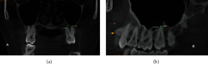
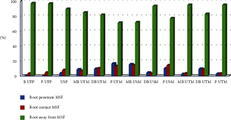
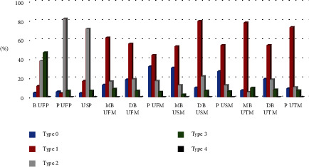
Materials and methods: This retrospective study evaluated 539 cone-beam computed tomography (CBCT) radiographs of patients over 20 years of age. Patients were divided into four groups according to age: group I (20-30 years), group II (31-40 years), group III (41-50 years), and group IV (more than 50 years). From coronal and sagittal images of CBCT, the vertical distance between the posterior maxillary root and the maxillary sinus was measured and classified according to its proximity to the maxillary sinus.
Results: Gender and size did not significantly affect the distance between maxillary posterior root and maxillary sinus. However, there was a significant increase in this distance with increased age. Mesiobuccal root of the second molar was the nearest root to the maxillary sinus (0.8 ± 1.62, p < 0.001), while the buccal root of the first premolar was the farthest root (5.39 ± 3.26, p < 0.001).
Conclusion: Regarding the population of this study, the buccal roots of the second molars are the closest to the sinus floor. Complications associated with maxillary molar extraction and implantation are greater at a younger age. Because the distance between posterior maxillary teeth and maxillary sinus was mostly type 1 (0-2 mm), clinicians are advised to perform CBCT to get a better understanding of the relationship between maxillary posterior roots and maxillary sinus before surgical intervention.
Copyright © 2022 Alaa Abdelqader Altaweel et al.
Conflict of interest statement
The authors declare that they have no conflicts of interest.
Figures
Similar articles
-
Morphometric analysis of the relationship between maxillary posterior teeth and maxillary sinus floor in central Indian population: A cone-beam computed tomography study.J Conserv Dent Endod. 2024 Apr;27(4):373-377. doi: 10.4103/JCDE.JCDE_353_23. Epub 2024 Apr 5. J Conserv Dent Endod. 2024. PMID: 38779206 Free PMC article.
-
Proximity of Posterior Teeth to the Maxillary Sinus and Buccal Bone Thickness: A Biometric Assessment Using Cone-beam Computed Tomography.J Endod. 2015 Nov;41(11):1839-46. doi: 10.1016/j.joen.2015.08.011. Epub 2015 Sep 26. J Endod. 2015. PMID: 26411520
-
Proximity of the roots of maxillary posterior teeth to the floor of maxillary sinus and cortical plate: A cone-beam computed tomography assessment.Indian J Dent Res. 2020 Nov-Dec;31(6):911-915. doi: 10.4103/ijdr.IJDR_871_18. Indian J Dent Res. 2020. PMID: 33753663
-
Evaluation of the relationship between maxillary posterior teeth and the maxillary sinus floor using cone-beam computed tomography.BMC Oral Health. 2018 Oct 3;18(1):164. doi: 10.1186/s12903-018-0626-z. BMC Oral Health. 2018. PMID: 30285721 Free PMC article.
-
Comparison of cone-beam computed tomography and panoramic imaging in assessing the relationship between posterior maxillary tooth roots and the maxillary sinus: A systematic review.J Investig Clin Dent. 2019 Aug;10(3):e12402. doi: 10.1111/jicd.12402. Epub 2019 Jan 28. J Investig Clin Dent. 2019. PMID: 30693662
Cited by
-
Evaluation of the Proximity of the Maxillary Teeth Root Apices to the Maxillary Sinus Floor in Romanian Subjects: A Cone-Beam Computed Tomography Study.Diagnostics (Basel). 2025 Jul 9;15(14):1741. doi: 10.3390/diagnostics15141741. Diagnostics (Basel). 2025. PMID: 40722490 Free PMC article.
-
Morphometric analysis of the relationship between maxillary posterior teeth and maxillary sinus floor in central Indian population: A cone-beam computed tomography study.J Conserv Dent Endod. 2024 Apr;27(4):373-377. doi: 10.4103/JCDE.JCDE_353_23. Epub 2024 Apr 5. J Conserv Dent Endod. 2024. PMID: 38779206 Free PMC article.
-
Endodontic Management of a Less Common Four-Rooted Maxillary Second Molar.Cureus. 2025 May 15;17(5):e84205. doi: 10.7759/cureus.84205. eCollection 2025 May. Cureus. 2025. PMID: 40525050 Free PMC article.
References
-
- Sharan A., Madjar D. Maxillary sinus pneumatization following extractions: a radiographic study. The International Journal of Oral & Maxillofacial Implants . 2008;23(1):48–56. - PubMed
-
- Lu Y., Liu Z., Zhang L. Associations between maxillary sinus mucosal thickening and apical periodontitis using cone-beam computed tomography scanning: a retrospective study. Journal of Endodontics . 2012;38(8):1069–1074. - PubMed
-
- Wehrbein H., Diedrich P. [Progressive pneumatization of the basal maxillary sinus after extraction and space closure] Fortschritte der Kieferorthopadie . 1992;53(2):p. 77 83. - PubMed
-
- Ariji Y., Obayashi N., Goto M. Roots of the maxillary first and second molars in horizontal relation to alveolar cortical plates and maxillary sinus: computed tomography assessment for infection spread. Clinical Oral Investigations . 2006;10(1):35–41. - PubMed
LinkOut - more resources
Full Text Sources
